Explore
 Validate
 Learn
LearnBS2155
antibody from Bioworld Technology, Inc
Targeting: ERAP1
A-LAP, ARTS-1, ERAAP1, KIAA0525, PILS-AP
 Western blot
 Immunocytochemistry
Antibody data
- Antibody Data
- Antigen structure
- References [0]
- Comments [0]
- Validations
- Western blot [1]
Submit
Validation data
Reference
Comment
Report error
- Product number
- BS2155 - Provider product page

- Provider
- Bioworld Technology, Inc
- Proper citation
- Bioworld Technology Cat#BS2155, RRID:AB_1663797
- Product name
- ERAP1 (K467) polyclonal antibody
- Antibody type
- Polyclonal
- Antigen
- Synthetic peptide, corresponding to amino acids 435-489 of Human ERAP1.
- Description
- The endoplasmic reticulum (ER) aminopeptidase 1 (ERAP1) is a 120 kDa protein localized to the lumen of the ER, which removes NH2-terminal residues from many antigenic precursors for MHC class I peptide presentation. Peptides that are presented by MHC class I on the surface of a cell must be 8-11 residues long, and ERAP1 specifically trims peptides of 9 amino acids or more. ERAP1 is also induced by interferon-ÎÛ. The gene encoding human ERAP1 maps to chromosome 5q15. ERAP1 has previously been characterized as adipocyte-derived leucine aminopeptidase (A-LAP), puromycin-insensitive leucine-specific aminopeptidase (PILS-AP) and aminopeptidase regulator of TNFR1 shedding (ARTS-1). A-LAP is thought to inactivate several bioactive peptides, including angiotensin II and, subsequently, may be involved in the regulation of blood pressure. PILS-AP is described as playing a role in angiogenesis by regulating the proliferation and migration of endothelial cells, and ARTS-1 is characterized as a TNFR1 binding protein that promotes TNFR1 shedding. Further research will be necessary to fully elucidate the functions of this protein.
- Reactivity
- Human, Mouse, Rat
- Host
- Rabbit
- Isotype
- IgG
- Vial size
- 100ul
- Concentration
- 1 mg/ml
- Storage
- Store at 4°C short term. Aliquot and store at -20°C long term. Avoid freeze-thaw cycles.
No comments: Submit comment
Supportive validation
- Submitted by
- Bioworld Technology, Inc (provider)
- Main image
- Experimental details
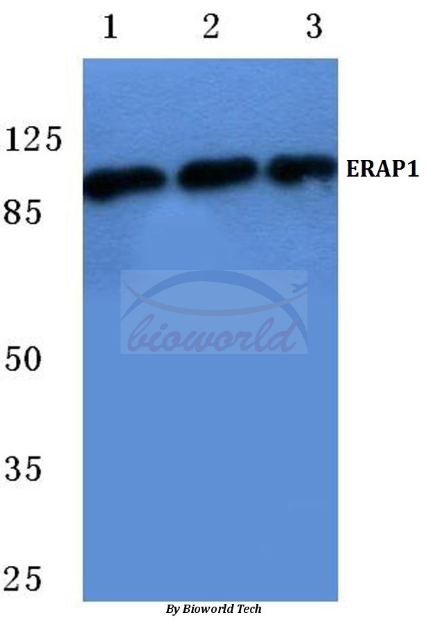
- Western blot (WB) analysis of ERAP1 (K467) pAb at 1:500 dilutionLane1:THP-1 cell lysateLane2:NIH-3T3 cell lysateLane3:Rat kidney tissue lysate