Explore
 Validate
 Learn
Learn Western blot
 Immunoprecipitation
Antibody data
- Antibody Data
- Antigen structure
- References [2]
- Comments [0]
- Validations
- Western blot [1]
- Immunocytochemistry [1]
- Immunohistochemistry [1]
Submit
Validation data
Reference
Comment
Report error
- Product number
- AF2334 - Provider product page

- Provider
- R&D Systems
- Product name
- Human Aminopeptidase PILS/ARTS1 Antibody
- Antibody type
- Polyclonal
- Description
- Antigen Affinity-purified. Detects human Aminopeptidase PILS/ARTS1 in direct ELISAs and Western blots. In direct ELISAs, approximately 45% cross-reactivity with recombinant mouse ARTS1 is observed.
- Reactivity
- Human
- Host
- Goat
- Conjugate
- Unconjugated
- Antigen sequence
EAW96077- Isotype
- IgG
- Vial size
- 100 ug
- Concentration
- LYOPH
- Storage
- Use a manual defrost freezer and avoid repeated freeze-thaw cycles. 12 months from date of receipt, -20 to -70 °C as supplied. 1 month, 2 to 8 °C under sterile conditions after reconstitution. 6 months, -20 to -70 °C under sterile conditions after reconstitution.
Submitted references Inositol-Requiring Enzyme 1-Mediated Downregulation of MicroRNA (miR)-146a and miR-155 in Primary Dermal Fibroblasts across Three TNFRSF1A Mutations Results in Hyperresponsiveness to Lipopolysaccharide.
The partial dissociation of MHC class I-bound peptides exposes their N terminus to trimming by endoplasmic reticulum aminopeptidase 1.
Harrison SR, Scambler T, Oubussad L, Wong C, Wittmann M, McDermott MF, Savic S
Frontiers in immunology 2018;9:173
Frontiers in immunology 2018;9:173
The partial dissociation of MHC class I-bound peptides exposes their N terminus to trimming by endoplasmic reticulum aminopeptidase 1.
Papakyriakou A, Reeves E, Beton M, Mikolajek H, Douglas L, Cooper G, Elliott T, Werner JM, James E
The Journal of biological chemistry 2018 May 18;293(20):7538-7548
The Journal of biological chemistry 2018 May 18;293(20):7538-7548
No comments: Submit comment
Supportive validation
- Submitted by
- R&D Systems (provider)
- Main image
- Experimental details
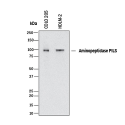
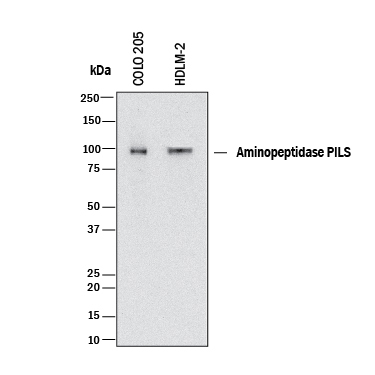
- Detection of Human Aminopeptidase PILS/ARTS1 by Western Blot. Western blot shows lysates of COLO 205 human colorectal adenocarcinoma cell line and HDLM-2 human Hodgkin's lymphoma cell line. PVDF membrane was probed with 1 µg/mL of Goat Anti-Human Aminopeptidase PILS/ARTS1 Antigen Affinity-purified Polyclonal Antibody (Catalog # AF2334) followed by HRP-conjugated Anti-Goat IgG Secondary Antibody (Catalog # HAF017). A specific band was detected for Aminopeptidase PILS/ARTS1 at approximately 100 kDa (as indicated). This experiment was conducted under reducing conditions and using Immunoblot Buffer Group 1.
Supportive validation
- Submitted by
- R&D Systems (provider)
- Main image

- Experimental details
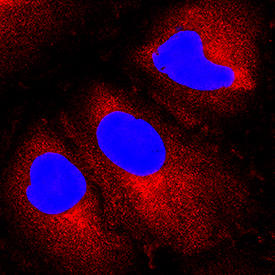
- Aminopeptidase PILS/ARTS1 in A549 Human Cell Line. Aminopeptidase PILS/ARTS1 was detected in immersion fixed A549 human lung carcinoma cell line using Goat Anti-Human Aminopeptidase PILS/ARTS1 Antigen Affinity-purified Polyclonal Antibody (Catalog # AF2334) at 15 µg/mL for 3 hours at room temperature. Cells were stained using the NorthernLights™ 557-conjugated Anti-Goat IgG Secondary Antibody (red; Catalog # NL001) and counterstained with DAPI (blue). Specific staining was localized to cytoplasm. View our protocol for Fluorescent ICC Staining of Cells on Coverslips.
Supportive validation
- Submitted by
- R&D Systems (provider)
- Main image
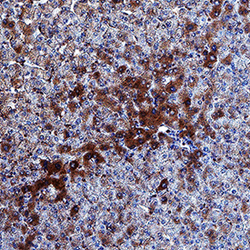
- Experimental details
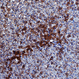
- Aminopeptidase PILS/ARTS1 in Human Liver. Aminopeptidase PILS/ARTS1 was detected in immersion fixed paraffin-embedded sections of human liver using Goat Anti-Human Aminopeptidase PILS/ARTS1 Antigen Affinity-purified Polyclonal Antibody (Catalog # AF2334) at 1 µg/mL overnight at 4 °C. Tissue was stained using the Anti-Goat HRP-DAB Cell & Tissue Staining Kit (brown; Catalog # CTS008) and counterstained with hematoxylin (blue). Specific staining was localized to hepatocyte cytoplasm. View our protocol for Chromogenic IHC Staining of Paraffin-embedded Tissue Sections.