Explore
 Validate
 Learn
Learn Western blot
 ELISA
ELISAAntibody data
- Antibody Data
- Antigen structure
- References [3]
- Comments [0]
- Validations
- Western blot [1]
- Immunocytochemistry [1]
- Immunohistochemistry [1]
- Flow cytometry [1]
Submit
Validation data
Reference
Comment
Report error
- Product number
- ABIN953208 - Provider product page

- Provider
- antibodies-online
- Product name
- anti-Low Density Lipoprotein Receptor-Related Protein 12 (LRP12) (C-Term) antibody
- Antibody type
- Polyclonal
- Antigen
- Other
- Reactivity
- Human
- Host
- Rabbit
- Vial size
- 0.1 mg
Submitted references Overexpression of LRP12, a gene contained within an 8q22 amplicon identified by high-resolution array CGH analysis of oral squamous cell carcinomas.
ST7 is a novel low-density lipoprotein receptor-related protein (LRP) with a cytoplasmic tail that interacts with proteins related to signal transduction pathways.
Cloning and characterization of a novel gene encoding a putative transmembrane protein with altered expression in some human transformed and tumor-derived cell lines.
Garnis C, Coe BP, Zhang L, Rosin MP, Lam WL
Oncogene 2004 Apr 1;23(14):2582-6
Oncogene 2004 Apr 1;23(14):2582-6
ST7 is a novel low-density lipoprotein receptor-related protein (LRP) with a cytoplasmic tail that interacts with proteins related to signal transduction pathways.
Battle MA, Maher VM, McCormick JJ
Biochemistry 2003 Jun 24;42(24):7270-82
Biochemistry 2003 Jun 24;42(24):7270-82
Cloning and characterization of a novel gene encoding a putative transmembrane protein with altered expression in some human transformed and tumor-derived cell lines.
Qing J, Wei D, Maher VM, McCormick JJ
Oncogene 1999 Jan 14;18(2):335-42
Oncogene 1999 Jan 14;18(2):335-42
No comments: Submit comment
Supportive validation
- Submitted by
- antibodies-online (provider)
- Main image
- Experimental details
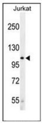
- Western blot analysis of LRP12 / ST7 Antibody (C-term) Cat.-No AP52525PU-N in Jurkat cell line lysates (35ug/lane). This demonstrates the LRP12 antibody detected the LRP12 protein (arrow).

Supportive validation
- Submitted by
- antibodies-online (provider)
- Main image
- Experimental details
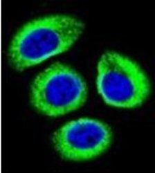
- Confocal immunofluorescent analysis of LRP12 / ST7 Antibody (C-term) Cat.-No AP52525PU-N with U-251MG cell followed by Alexa Fluor 488-conjugated goat anti-rabbit lgG (green). DAPI was used to stain the cell nuclear (blue).

Supportive validation
- Submitted by
- antibodies-online (provider)
- Main image

- Experimental details
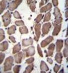
- Immunohistochemistry analysis in formalin fixed and paraffin embedded human skeletal muscle reacted with LRP12 / ST7 Antibody (C-term) Cat.-No AP52525PU-N, which was peroxidase conjugated to the secondary antibody and DAB staining.
Supportive validation
- Submitted by
- antibodies-online (provider)
- Main image

- Experimental details
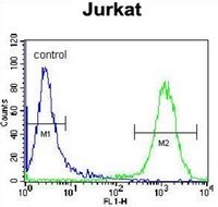
- Flow cytometric analysis of Jurkat cells using LRP12 / ST7 Antibody (C-term) Cat.-No AP52525PU-N (right histogram) compared to a negative control cell (left histogram). FITC-conjugated donkey-anti-rabbit secondary antibodies were used for the analysis.