Explore
 Validate
 Learn
Learn Western blot
 Immunohistochemistry
Antibody data
- Antibody Data
- Antigen structure
- References [1]
- Comments [0]
- Validations
- Immunohistochemistry [1]
- Other assay [2]
Submit
Validation data
Reference
Comment
Report error
- Product number
- PA5-32043 - Provider product page

- Provider
- Invitrogen Antibodies
- Product name
- LACTB2 Polyclonal Antibody
- Antibody type
- Polyclonal
- Antigen
- Recombinant full-length protein
- Description
- Recommended positive controls: A549, HCT116. Predicted reactivity: Mouse (82%), Rat (81%), Pig (86%), Bovine (89%). Store product as a concentrated solution. Centrifuge briefly prior to opening the vial.
- Reactivity
- Human
- Host
- Rabbit
- Isotype
- IgG
- Vial size
- 100 μL
- Concentration
- 0.61 mg/mL
- Storage
- Store at 4°C short term. For long term storage, store at -20°C, avoiding freeze/thaw cycles.
Submitted references Identification of LACTB2, a metallo-β-lactamase protein, as a human mitochondrial endoribonuclease.
Levy S, Allerston CK, Liveanu V, Habib MR, Gileadi O, Schuster G
Nucleic acids research 2016 Feb 29;44(4):1813-32
Nucleic acids research 2016 Feb 29;44(4):1813-32
No comments: Submit comment
Supportive validation
- Submitted by
- Invitrogen Antibodies (provider)
- Main image
- Experimental details
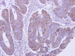
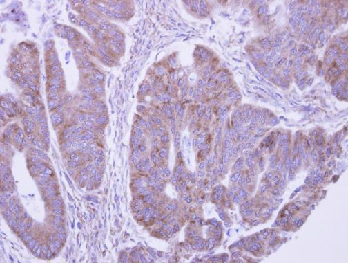
- Immunohistochemical analysis of paraffin-embedded human colon carcinoma, using LACTB2 Polyclonal Antibody (Product # PA5-32043) antibody at 1:500 dilution. Antigen Retrieval: EDTA based buffer, pH 8.0, 15 min.
Supportive validation
- Submitted by
- Invitrogen Antibodies (provider)
- Main image

- Experimental details
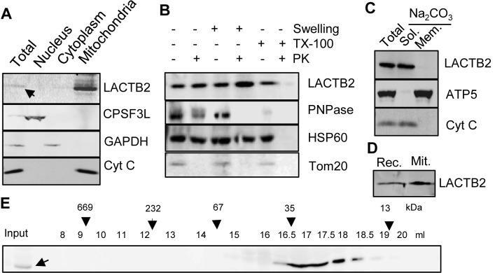
- Figure 1. LACTB2 is a mitochondrial, soluble and monomeric protein. ( A ) Mitochondria localization. HeLa cells were disrupted and the nuclear, cytoplasmic and mitochondria-containing fractions were separated and analyzed for the localization of LACTB2, using an immunoblotting assay. The nucleus-located protein, CPSF3L, was used as marker for nuclear proteins. Glyceraldehyde-3-phosphate dehydrogenase (GAPDH) served as a cytosolic marker and cytochrome C (Cyt C) as a mitochondrial marker. ( B ) Immunoblot analysis of mitochondrial extract following Proteinase K accessibility assay. TX-100, Triton X-100. PK, Proteinase K. PNPase is a mitochondrial protein located mainly in the intermembrane space and a small amount in the matrix. HSP60 is a mitochondrial matrix protein. Tom20 is a mitochondrial outer membrane protein. ( C ) LACTB2 is a soluble protein. Immunoblotting analysis of mitochondrial extract following alkaline sodium carbonate (Na 2 CO 3 ) extraction of soluble and peripheral membrane proteins. T, total extract. S, supernatant. P, pellet. Subunit 5 of the ATP synthase (ATP5) was used as a marker for intrinsic membrane proteins and Cyt C as a marker of soluble proteins. (D) Immunoblot analysis of the recombinant LACTB2 (Rec.) and the native protein in isolated mitochondria (Mit.) showing the same size on SDS-PAGE. Therefore, the mitochondria targeting signal is not cleaved upon entering the mitochondria and remain an integral part of the mature protein. ( E ) LACTB2 is
- Submitted by
- Invitrogen Antibodies (provider)
- Main image
- Experimental details
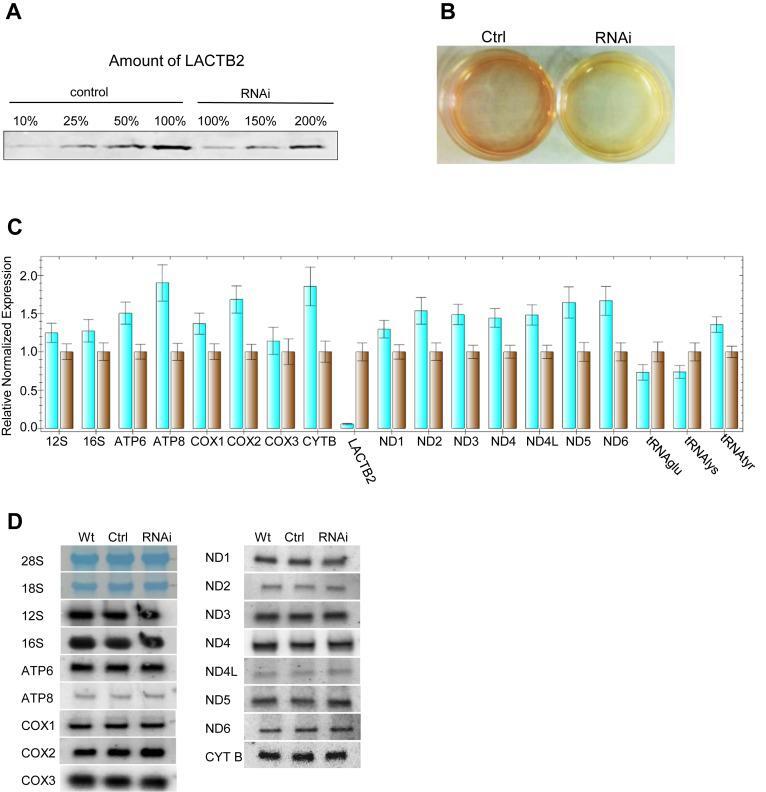
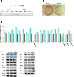
- Figure 8. Downregulation of LACTB2 expression resulted in modest accumulation of mitochondrial transcripts. ( A ) Quantitation of the amount of LACTB2 in the siRNA treated cells. siRNA duplex targeted for LACTB2 or scrambled siRNAs duplexes, as a negative control, were transfected into HEK-293 cells. Cells were collected 48 h post-transfection and the amount of LACTB2 was determined using immunoblotting assay. Proteins from equal number of cells were loaded in the lanes labeled 100%. Proteins from 50, 25 and 10% of the control and 150 and 200% of the siRNA LACTB2 treated cells were analyzed for the amount of LACTB2, in order to determine the degree of down-expression. ( B ) Medium acidification due to treatment with siRNA duplex, 48 h post-transfection. Ctrl--cells treated with scrambled siRNAs duplexes as a negative control. RNAi--cells treated with siRNA duplex targeted to LACTB2. ( C ) The amount of mitochondrial transcripts in the cells treated with the siRNA duplex targeted to LACTB2 (turquoise bars), or with scrambled siRNAs duplexes as a negative control (brown bars), was analyzed by qRT-PCR. The GAPDH transcript was used as the reference gene for normalization of the expression. The error bars display the standard errors of at least three independent experiments. ( D ) Northern blot analysis of HEK-293 cells that were not transfected (WT), transfected with scrambled siRNAs as control (Ctrl) or transfected with LACTB2 siRNA (RNAi). Hybridizations were performed with pr