Explore
 Validate
 Learn
Learn Western blot
 Immunocytochemistry
Antibody data
- Antibody Data
- Antigen structure
- References [28]
- Comments [0]
- Validations
- Western blot [1]
- Immunocytochemistry [1]
Submit
Validation data
Reference
Comment
Report error
- Product number
- HPA045910 - Provider product page

- Provider
- Atlas Antibodies
- Proper citation
- Atlas Antibodies Cat#HPA045910, RRID:AB_10960409
- Product name
- Anti-CRBN
- Antibody type
- Polyclonal
- Description
- Polyclonal Antibody against Human CRBN, Gene description: cereblon, Alternative Gene Names: MRT2, MRT2A, Validated applications: ICC, WB, Uniprot ID: Q96SW2, Storage: Store at +4°C for short term storage. Long time storage is recommended at -20°C.
- Reactivity
- Human, Mouse, Rat
- Host
- Rabbit
- Conjugate
- Unconjugated
- Isotype
- IgG
- Vial size
- 100 µl
- Concentration
- 0.2 mg/ml
- Storage
- Store at +4°C for short term storage. Long time storage is recommended at -20°C.
- Handling
- The antibody solution should be gently mixed before use.
Submitted references A Tandem-Affinity Purification Method for Identification of Primary Intracellular Drug-Binding Proteins
Activity-based profiling of cullin–RING E3 networks by conformation-specific probes
Direct-to-biology, automated, nano-scale synthesis, and phenotypic screening-enabled E3 ligase modulator discovery
Sec61 blockade therapy overrides resistance to proteasome inhibitors and immunomodulatory drugs in multiple myeloma
A Novel BRD Family PROTAC Inhibitor dBET1 Exerts Great Anti-Cancer Effects by Targeting c-MYC in Acute Myeloid Leukemia Cells
The BRD4 Inhibitor dBET57 Exerts Anticancer Effects by Targeting Superenhancer-Related Genes in Neuroblastoma
Profiling of diverse tumor types establishes the broad utility of VHL-based ProTaCs and triages candidate ubiquitin ligases.
Cereblon Regulates the Proteotoxicity of Tau by Tuning the Chaperone Activity of DNAJA1
BRD4 PROTAC degrader ARV-825 inhibits T-cell acute lymphoblastic leukemia by targeting 'Undruggable' Myc-pathway genes
Pomalidomide restores immune recognition of primary effusion lymphoma through upregulation of ICAM-1 and B7-2
Regulation of AMPK Activity by CRBN Is Independent of the Thalidomide-CRL4CRBN Protein Degradation Axis
Immunomodulatory effect of NEDD8-activating enzyme inhibition in Multiple Myeloma: upregulation of NKG2D ligands and sensitization to Natural Killer cell recognition
ARV-825 Demonstrates Antitumor Activity in Gastric Cancer via MYC-Targets and G2M-Checkpoint Signaling Pathways
Long-term depletion of cereblon induces mitochondrial dysfunction in cancer cells.
The CDK inhibitor CR8 acts as a molecular glue degrader that depletes cyclin K
Role of cereblon in angiogenesis and in mediating the antiangiogenic activity of immunomodulatory drugs
Crbn modulates calcium influx by regulating Orai1 during efferocytosis
PROTAC Bromodomain Inhibitor ARV-825 Displays Anti-Tumor Activity in Neuroblastoma by Repressing Expression of MYCN or c-Myc
Identification of lenalidomide resistance pathways in myeloma and targeted resensitization using cereblon replacement, inhibition of STAT3 or targeting of IRF4
Disordered region of cereblon is required for efficient degradation by proteolysis-targeting chimera
RUNX proteins desensitize multiple myeloma to lenalidomide via protecting IKZFs from degradation
The homeobox transcription factor MEIS2 is a regulator of cancer cell survival and IMiDs activity in Multiple Myeloma: modulation by Bromodomain and Extra-Terminal (BET) protein inhibitors
Dual inhibition of DNMTs and EZH2 can overcome both intrinsic and acquired resistance of myeloma cells to IMiDs in a cereblon‐independent manner
p97/VCP promotes degradation of CRBN substrate glutamine synthetase and neosubstrates
Amyloid Precursor Protein (APP) May Act as a Substrate and a Recognition Unit for CRL4CRBN and Stub1 E3 Ligases Facilitating Ubiquitination of Proteins Involved in Presynaptic Functions and Neurodegeneration
Rabex-5 is a lenalidomide target molecule that negatively regulates TLR-induced type 1 IFN production
Measuring cereblon as a biomarker of response or resistance to lenalidomide and pomalidomide requires use of standardized reagents and understanding of gene complexity
Islam S, Gour J, Beer T, Tang H, Cassel J, Salvino J, Busino L
ACS Chemical Biology 2024;19(2):233-242
ACS Chemical Biology 2024;19(2):233-242
Maddineni A, Liang Z, Jambardi S, Roy S, Tycko J, Patil A, Manzano M, Gottwein E
2024
2024
Activity-based profiling of cullin–RING E3 networks by conformation-specific probes
Henneberg L, Singh J, Duda D, Baek K, Yanishevski D, Murray P, Mann M, Sidhu S, Schulman B
Nature Chemical Biology 2023;19(12):1513-1523
Nature Chemical Biology 2023;19(12):1513-1523
Direct-to-biology, automated, nano-scale synthesis, and phenotypic screening-enabled E3 ligase modulator discovery
Wang Z, Shaabani S, Gao X, Ng Y, Sapozhnikova V, Mertins P, Krönke J, Dömling A
Nature Communications 2023;14(1)
Nature Communications 2023;14(1)
Sec61 blockade therapy overrides resistance to proteasome inhibitors and immunomodulatory drugs in multiple myeloma
Domenger A, Ricci D, Mayau V, Majlessi L, Marcireau C, Dadaglio G, Demangel C
Frontiers in Oncology 2023;13
Frontiers in Oncology 2023;13
A Novel BRD Family PROTAC Inhibitor dBET1 Exerts Great Anti-Cancer Effects by Targeting c-MYC in Acute Myeloid Leukemia Cells
Zhang K, Gao L, Wang J, Chu X, Zhang Z, Zhang Y, Fang F, Tao Y, Li X, Tian Y, Li Z, Sang X, Ma L, Lu L, Chen Y, Yu J, Zhuo R, Wu S, Pan J, Hu S
Pathology and Oncology Research 2022;28
Pathology and Oncology Research 2022;28
The BRD4 Inhibitor dBET57 Exerts Anticancer Effects by Targeting Superenhancer-Related Genes in Neuroblastoma
Jia S, Zhuo R, Zhang Z, Yang Y, Tao Y, Wang J, Li X, Xie Y, Li G, Wu D, Chen Y, Yu J, Feng C, Li Z, Zhou R, Yang R, Yang P, Zhou B, Wan X, Wu Y, Jiao W, Zhou N, Fang F, Pan J, Zhong J
Journal of Immunology Research 2022;2022
Journal of Immunology Research 2022;2022
Profiling of diverse tumor types establishes the broad utility of VHL-based ProTaCs and triages candidate ubiquitin ligases.
Luo X, Archibeque I, Dellamaggiore K, Smither K, Homann O, Lipford JR, Mohl D
iScience 2022 Mar 18;25(3):103985
iScience 2022 Mar 18;25(3):103985
Cereblon Regulates the Proteotoxicity of Tau by Tuning the Chaperone Activity of DNAJA1
Akber U, Jo H, Jeon S, Yang S, Bong S, Lim S, Kim Y, Park Z, Park C
The Journal of Neuroscience 2021;41(24):5138-5156
The Journal of Neuroscience 2021;41(24):5138-5156
BRD4 PROTAC degrader ARV-825 inhibits T-cell acute lymphoblastic leukemia by targeting 'Undruggable' Myc-pathway genes
Wu S, Jiang Y, Hong Y, Chu X, Zhang Z, Tao Y, Fan Z, Bai Z, Li X, Chen Y, Li Z, Ding X, Lv H, Du X, Lim S, Zhang Y, Huang S, Lu J, Pan J, Hu S
Cancer Cell International 2021;21(1)
Cancer Cell International 2021;21(1)
Pomalidomide restores immune recognition of primary effusion lymphoma through upregulation of ICAM-1 and B7-2
Lieberman P, Shrestha P, Davis D, Jaeger H, Stream A, Aisabor A, Yarchoan R
PLOS Pathogens 2021;17(1):e1009091
PLOS Pathogens 2021;17(1):e1009091
Regulation of AMPK Activity by CRBN Is Independent of the Thalidomide-CRL4CRBN Protein Degradation Axis
Yang S, Jeon S, Baek J, Lee K, Park C
Pharmaceuticals 2021;14(6):512
Pharmaceuticals 2021;14(6):512
Immunomodulatory effect of NEDD8-activating enzyme inhibition in Multiple Myeloma: upregulation of NKG2D ligands and sensitization to Natural Killer cell recognition
Petillo S, Capuano C, Molfetta R, Fionda C, Mekhloufi A, Pighi C, Antonangeli F, Zingoni A, Soriani A, Petrucci M, Galandrini R, Paolini R, Santoni A, Cippitelli M
Cell Death & Disease 2021;12(9)
Cell Death & Disease 2021;12(9)
ARV-825 Demonstrates Antitumor Activity in Gastric Cancer via MYC-Targets and G2M-Checkpoint Signaling Pathways
Liao X, Qian X, Zhang Z, Tao Y, Li Z, Zhang Q, Liang H, Li X, Xie Y, Zhuo R, Chen Y, Jiang Y, Cao H, Niu J, Xue C, Ni J, Pan J, Cui D
Frontiers in Oncology 2021;11
Frontiers in Oncology 2021;11
Long-term depletion of cereblon induces mitochondrial dysfunction in cancer cells.
Park S, Kim K, Haam K, Ban HS, Kim JA, Park BC, Park SG, Kim S, Kim JH
BMB reports 2021 Jun;54(6):305-310
BMB reports 2021 Jun;54(6):305-310
The CDK inhibitor CR8 acts as a molecular glue degrader that depletes cyclin K
Słabicki M, Kozicka Z, Petzold G, Li Y, Manojkumar M, Bunker R, Donovan K, Sievers Q, Koeppel J, Suchyta D, Sperling A, Fink E, Gasser J, Wang L, Corsello S, Sellar R, Jan M, Gillingham D, Scholl C, Fröhling S, Golub T, Fischer E, Thomä N, Ebert B
Nature 2020;585(7824):293-297
Nature 2020;585(7824):293-297
Role of cereblon in angiogenesis and in mediating the antiangiogenic activity of immunomodulatory drugs
Beedie S, Huang P, Harris E, Strope J, Mahony C, Chau C, Vargesson N, Figg W
The FASEB Journal 2020;34(9):11395-11404
The FASEB Journal 2020;34(9):11395-11404
Crbn modulates calcium influx by regulating Orai1 during efferocytosis
Moon H, Min C, Kim G, Kim D, Kim K, Lee S, Moon B, Yang S, Lee J, Yang S, Cho S, Lee G, Lee C, Park C, Park D
Nature Communications 2020;11(1)
Nature Communications 2020;11(1)
PROTAC Bromodomain Inhibitor ARV-825 Displays Anti-Tumor Activity in Neuroblastoma by Repressing Expression of MYCN or c-Myc
Li Z, Lim S, Tao Y, Li X, Xie Y, Yang C, Zhang Z, Jiang Y, Zhang X, Cao X, Wang H, Qian G, Wu Y, Li M, Fang F, Liu Y, Fu M, Ding X, Zhu Z, Lv H, Lu J, Xiao S, Hu S, Pan J
Frontiers in Oncology 2020;10
Frontiers in Oncology 2020;10
Identification of lenalidomide resistance pathways in myeloma and targeted resensitization using cereblon replacement, inhibition of STAT3 or targeting of IRF4
Zhu Y, Shi C, Bruins L, Wang X, Riggs D, Porter B, Ahmann J, de Campos C, Braggio E, Bergsagel P, Stewart A
Blood Cancer Journal 2019;9(2)
Blood Cancer Journal 2019;9(2)
Disordered region of cereblon is required for efficient degradation by proteolysis-targeting chimera
Kim K, Lee D, Park S, Jo S, Ku B, Park S, Park B, Jeon Y, Ahn S, Kang C, Hwang D, Chae S, Ha J, Kim S, Hwang J, Kim J
Scientific Reports 2019;9(1)
Scientific Reports 2019;9(1)
RUNX proteins desensitize multiple myeloma to lenalidomide via protecting IKZFs from degradation
Zhou N, Gutierrez-Uzquiza A, Zheng X, Chang R, Vogl D, Garfall A, Bernabei L, Saraf A, Florens L, Washburn M, Illendula A, Bushweller J, Busino L
Leukemia 2019;33(8):2006-2021
Leukemia 2019;33(8):2006-2021
The homeobox transcription factor MEIS2 is a regulator of cancer cell survival and IMiDs activity in Multiple Myeloma: modulation by Bromodomain and Extra-Terminal (BET) protein inhibitors
Abruzzese M, Bilotta M, Fionda C, Zingoni A, Soriani A, Petrucci M, Ricciardi M, Molfetta R, Paolini R, Santoni A, Cippitelli M
Cell Death & Disease 2019;10(4)
Cell Death & Disease 2019;10(4)
Dual inhibition of DNMTs and EZH2 can overcome both intrinsic and acquired resistance of myeloma cells to IMiDs in a cereblon‐independent manner
Dimopoulos K, Søgaard Helbo A, Fibiger Munch‐Petersen H, Sjö L, Christensen J, Sommer Kristensen L, Asmar F, Hermansen N, O'Connel C, Gimsing P, Liang G, Grønbæk K
Molecular Oncology 2017;12(2):180-195
Molecular Oncology 2017;12(2):180-195
p97/VCP promotes degradation of CRBN substrate glutamine synthetase and neosubstrates
Nguyen T, Li J, Lu C, Mamrosh J, Lu G, Cathers B, Deshaies R
Proceedings of the National Academy of Sciences 2017;114(14):3565-3571
Proceedings of the National Academy of Sciences 2017;114(14):3565-3571
Amyloid Precursor Protein (APP) May Act as a Substrate and a Recognition Unit for CRL4CRBN and Stub1 E3 Ligases Facilitating Ubiquitination of Proteins Involved in Presynaptic Functions and Neurodegeneration
Del Prete D, Rice R, Rajadhyaksha A, D'Adamio L
Journal of Biological Chemistry 2016;291(33):17209-17227
Journal of Biological Chemistry 2016;291(33):17209-17227
Rabex-5 is a lenalidomide target molecule that negatively regulates TLR-induced type 1 IFN production
Millrine D, Tei M, Gemechu Y, Kishimoto T
Proceedings of the National Academy of Sciences 2016;113(38):10625-10630
Proceedings of the National Academy of Sciences 2016;113(38):10625-10630
Measuring cereblon as a biomarker of response or resistance to lenalidomide and pomalidomide requires use of standardized reagents and understanding of gene complexity
Gandhi A, Mendy D, Waldman M, Chen G, Rychak E, Miller K, Gaidarova S, Ren Y, Wang M, Breider M, Carmel G, Mahmoudi A, Jackson P, Abbasian M, Cathers B, Schafer P, Daniel T, Lopez‐Girona A, Thakurta A, Chopra R
British Journal of Haematology 2013;164(2):233-244
British Journal of Haematology 2013;164(2):233-244
No comments: Submit comment
Enhanced validation
- Submitted by
- Atlas Antibodies (provider)
- Enhanced method
- Genetic validation
- Main image
- Experimental details
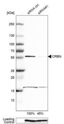
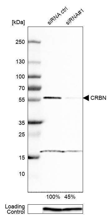
- Western blot analysis in HEK293 cells transfected with control siRNA, target specific siRNA probe #1, using Anti-CRBN antibody. Remaining relative intensity is presented. Loading control: Anti-GAPDH.
- Sample type
- Human
- Protocol
- Protocol
Supportive validation
- Submitted by
- Atlas Antibodies (provider)
- Main image

- Experimental details
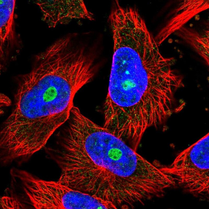
- Immunofluorescent staining of human cell line U-251 MG shows localization to nucleoli.
- Sample type
- Human