Explore
 Validate
 Learn
Learn Western blot
Antibody data
- Antibody Data
- Antigen structure
- References [0]
- Comments [0]
- Validations
- Western blot [1]
- Immunohistochemistry [2]
Submit
Validation data
Reference
Comment
Report error
- Product number
- ASC-007-200UL - Provider product page

- Provider
- Invitrogen Antibodies
- Product name
- SCN2B (NaV beta 2) Polyclonal Antibody
- Antibody type
- Polyclonal
- Antigen
- Other
- Reactivity
- Human, Mouse, Rat
- Host
- Rabbit
- Isotype
- IgG
- Vial size
- 200 µL
- Concentration
- 0.75 mg/mL
- Storage
- -20° C, Avoid Freeze/Thaw Cycles
No comments: Submit comment
Supportive validation
- Submitted by
- Invitrogen Antibodies (provider)
- Main image
- Experimental details
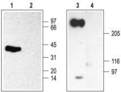
- Western blot analysis of rat brain membranes under reducing (lanes 1 and 2) or non-reducing (lanes 3 and 4) conditions:* - 1,3. Anti-SCN2B (NaV beta 2) Antibody (#ASC-007), (1:200).2,4. Anti-SCN2B (NaV beta 2) Antibody , preincubated with SCN2B/Nav beta 2 Blocking Peptide (#BLP-SC007).*NaV beta 2is disulfide-linked to the alpha subunit of voltage-gated Na+channels (Schmidt, J.W. and Catterall, W.A. (1986)Cell46,437.).

Supportive validation
- Submitted by
- Invitrogen Antibodies (provider)
- Main image

- Experimental details
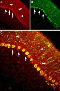
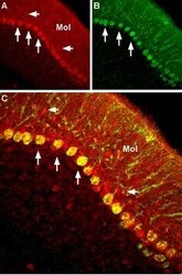
- Expression of NaV beta 2in rat cerebellum - Immunohistochemical staining of rat cerebellum using Anti-SCN2B (NaV beta 2) Antibody (#ASC-007). A. NaV beta 2channel (red) appears in Purkinje cells (horizontal arrows) and is distributed diffusely in the molecular layer (Mol). B. Staining of Purkinje nerve cells with mouse Anti-calcium binding protein (CBD28k, green) demonstrates the restriction of NaV beta 2to cell bodies. C. Confocal merge of NaV beta 2and CBD28k.
- Submitted by
- Invitrogen Antibodies (provider)
- Main image
- Experimental details
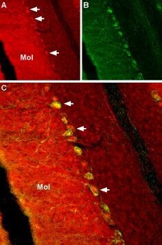
- Expression of NaV beta 2in mouse cerebellum - Immunohistochemical staining of mouse cerebellum using Anti-SCN2B (NaV beta 2) Antibody (#ASC-007). A. NaV beta 2channel (red) appears in Purkinje cells (vertical arrows) and in small cells inthe molecular layer (horizontal arrows, Mol). B. Staining of Purkinje nerve cells with mouse Anti-calcium binding protein (CBD28k, green) demonstrates the restriction of NaV beta 2to cell bodies. C. Confocal merge of NaV beta 2and CBD28k.