Explore
 Validate
 Learn
Learn Western blot
Antibody data
- Antibody Data
- Antigen structure
- References [0]
- Comments [0]
- Validations
- Western blot [2]
- Immunocytochemistry [1]
Submit
Validation data
Reference
Comment
Report error
- Product number
- MAB6195 - Provider product page

- Provider
- R&D Systems
- Product name
- Human CHD1 Antibody
- Antibody type
- Monoclonal
- Description
- Protein A or G purified from hybridoma culture supernatant. Detects human CHD1 in direct ELISAs and Western blots. In direct ELISAs, no cross-reactivity with recombinant human CHD1L, 5, or 7 is observed.
- Reactivity
- Human
- Host
- Mouse
- Conjugate
- Unconjugated
- Antigen sequence
O14646- Isotype
- IgG
- Antibody clone number
- 677616
- Vial size
- 100 ug
- Concentration
- LYOPH
- Storage
- Use a manual defrost freezer and avoid repeated freeze-thaw cycles. 12 months from date of receipt, -20 to -70 °C as supplied. 1 month, 2 to 8 °C under sterile conditions after reconstitution. 6 months, -20 to -70 °C under sterile conditions after reconstitution.
No comments: Submit comment
Supportive validation
- Submitted by
- R&D Systems (provider)
- Main image
- Experimental details
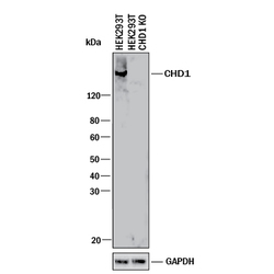
- Western Blot Shows Human CHD1 Specificity by Using Knockout Cell Line. Western blot shows lysates of HEK293T human embryonic kidney parental cell line and CHD1 knockout HEK293T cell line (KO). PVDF membrane was probed with 0.5 µg/mL of Mouse Anti-Human CHD1 Monoclonal Antibody (Catalog # MAB6195) followed by HRP-conjugated Anti-Mouse IgG Secondary Antibody (Catalog # HAF018). A specific band was detected for CHD1 at approximately 210 kDa (as indicated) in the parental HEK293T cell line, but is not detectable in knockout HEK293T cell line. GAPDH (Catalog # MAB5718) is shown as a loading control. This experiment was conducted under reducing conditions and using Immunoblot Buffer Group 1.

- Submitted by
- R&D Systems (provider)
- Main image
- Experimental details
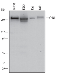
- Detection of Human and Mouse CHD1 by Western Blot. Western blot shows lysates of Jurkat human acute T cell leukemia cell line, K562 human chronic myelogenous leukemia cell line, Raji human Burkitt's lymphoma cell line, and BaF3 mouse pro-B cell line. PVDF Membrane was probed with 0.5 µg/mL of Human CHD1 Monoclonal Antibody (Catalog # MAB6195) followed by HRP-conjugated Anti-Mouse IgG Secondary Antibody (Catalog # HAF007). A specific band was detected for CHD1 at approximately 220 kDa (as indicated). This experiment was conducted under reducing conditions and using Immunoblot Buffer Group 1.

Supportive validation
- Submitted by
- R&D Systems (provider)
- Main image

- Experimental details
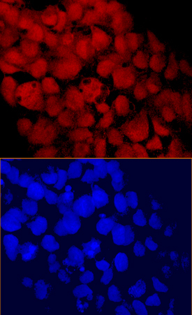
- CHD1 in BG01V Human Stem Cells. CHD1 was detected in immersion fixed BG01V human embryonic stem cells using Human CHD1 Monoclonal Antibody (Catalog # MAB6195) at 10 µg/mL for 3 hours at room temperature. Cells were stained using the NorthernLights™ 557-conjugated Anti-Mouse IgG Secondary Antibody (red, upper panel; Catalog # NL007) and counterstained with DAPI (blue, lower panel). Specific staining was localized to nuclei. View our protocol for Fluorescent ICC Staining of Cells on Coverslips.