 Explore
 Validate
 Learn
Learn Western blot
Antibody data
- Antibody Data
- Antigen structure
- References [3]
- Comments [0]
- Validations
- Western blot [2]
- Immunocytochemistry [6]
Submit
Validation data
Reference
Comment
Report error
- Product number
- PA1-1069 - Provider product page

- Provider
- Invitrogen Antibodies
- Product name
- TGN46 Polyclonal Antibody
- Antibody type
- Polyclonal
- Antigen
- Synthetic peptide
- Description
- PA1-1069 detects TGN46 in human samples. PA1-1069 has been successfully used in Western blot and immunofluorescence procedures. By Western blot, this antibody detects a ~51 kDa protein representing TGN46 in HEK293 and HeLa cell lysates. The PA1-1069 immunogen is a synthetic peptide corresponding to residues V(252) V P E Q P S W K D H S K P I S(267) of human TGN46. This peptide (Cat. # PEP-291) is available for use in neutralization and control experiments.
- Reactivity
- Human
- Host
- Rabbit
- Isotype
- IgG
- Vial size
- 100 µg
- Concentration
- 1 mg/mL
- Storage
- -20° C, Avoid Freeze/Thaw Cycles
Submitted references Influenza A Virus Infection Activates NLRP3 Inflammasome through Trans-Golgi Network Dispersion.
Human plasma C3 is essential for the development of memory B, but not T, lymphocytes.
Quantitative proteomic analysis of PCSK9 gain of function in human hepatic HuH7 cells.
Pandey KP, Zhou Y
Viruses 2022 Jan 5;14(1)
Viruses 2022 Jan 5;14(1)
Human plasma C3 is essential for the development of memory B, but not T, lymphocytes.
Jiménez-Reinoso A, Marin AV, Subias M, López-Lera A, Román-Ortiz E, Payne K, Ma CS, Arbore G, Kolev M, Freeley SJ, Kemper C, Tangye SG, Fernández-Malavé E, Rodríguez de Córdoba S, López-Trascasa M, Regueiro JR
The Journal of allergy and clinical immunology 2018 Mar;141(3):1151-1154.e14
The Journal of allergy and clinical immunology 2018 Mar;141(3):1151-1154.e14
Quantitative proteomic analysis of PCSK9 gain of function in human hepatic HuH7 cells.
Denis N, Palmer-Smith H, Elisma F, Busuttil A, Wright TG, Bou Khalil M, Prat A, Seidah NG, Chrétien M, Mayne J, Figeys D
Journal of proteome research 2011 Apr 1;10(4):2011-26
Journal of proteome research 2011 Apr 1;10(4):2011-26
No comments: Submit comment
Supportive validation
- Submitted by
- Invitrogen Antibodies (provider)
- Main image
- Experimental details
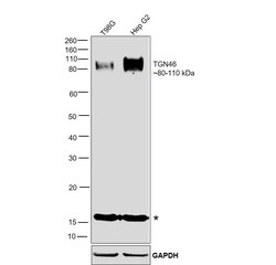
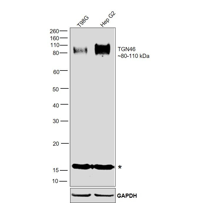
- Western blot was performed using Anti-TGN46 Polyclonal Antibody (Product # PA1-1069) and an 80-110 kDa band corresponding to Tgoln2 was observed across all the cell lines tested. A non-specific band (*) was seen above 15 kDa. Whole-cell extracts (30 µg lysate) of T98G (Lane 1), and Hep G2 (Lane 2) were electrophoresed using NuPAGE™ 4-12% Bis-Tris Protein Gel (Product # NP0321BOX). Resolved proteins were then transferred onto a nitrocellulose membrane (Product # IB23001) by iBlot® 2 Dry Blotting System (Product # IB21001). The blot was probed with the primary antibody (1:500 dilution) and detected by chemiluminescence with Goat anti-Rabbit IgG (H+L) Superclonal™ Recombinant Secondary Antibody, HRP (Product # A27036,1:20,000 dilution) using the iBright™ FL1500 Imaging System (Product # A44115). Chemiluminescent detection was performed using SuperSignal™ West Pico PLUS Chemiluminescent Substrate (Product # 34580).
- Submitted by
- Invitrogen Antibodies (provider)
- Main image
- Experimental details
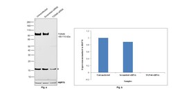
- Knockdown of Tgoln2 was achieved by transfecting T98G with Tgoln2-specific siRNAs (Silencer® select Product # S20855, S20854). Western blot analysis (Fig. a) was performed using Whole cell extracts from the Tgoln2 knockdown cells (lane 3), non-targeting scrambled siRNA transfected cells (lane 2), and untransfected cells (lane 1). The blot was probed with TGN46 Polyclonal Antibody (Product # PA1-1069, 1:500 dilution) and Goat anti-Rabbit IgG (H+L) Superclonal™ Recombinant Secondary Antibody, HRP (Product # A27036, 1:20,000 dilution). Densitometric analysis of this western blot is shown in the histogram (Fig. b). A decrease in signal upon siRNA-mediated knockdown confirms that the antibody is specific to Tgoln2.

Supportive validation
- Submitted by
- Invitrogen Antibodies (provider)
- Main image
- Experimental details
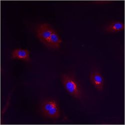
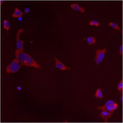
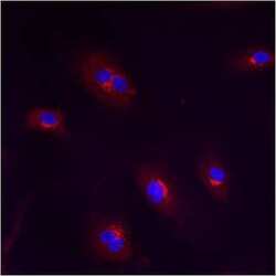
- Immunofluorescent analysis of Trans Golgi Network 46 using anti-Trans Golgi Network 46 polyclonal antibody (Product # PA1-1069) shows staining in HMVEC Cells.

- Submitted by
- Invitrogen Antibodies (provider)
- Main image
- Experimental details
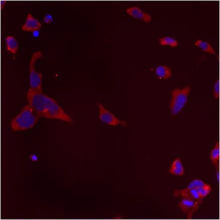
- Immunofluorescent analysis of Trans Golgi Network 46 using anti-Trans Golgi Network 46 polyclonal antibody (Product # PA1-1069) shows staining in p19 Cells.
- Submitted by
- Invitrogen Antibodies (provider)
- Main image
- Experimental details
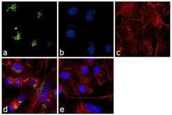
- Immunofluorescence analysis of Trans-Golgi Network 46 was performed using 70% confluent log phase HepG2 cells. The cells were fixed with 4% paraformaldehyde for 10 minutes, permeabilized with 0.1% Triton™ X-100 for 10 minutes, and blocked with 1% BSA for 1 hour at room temperature. The cells were labeled with Trans-Golgi Network 46 Rabbit Polyclonal Antibody (Product # PA1-1069) at 2µg/mLin 0.1% BSA, incubated for 3 hours at room temperature and then labeled with Goat anti-Rabbit IgG (H+L) Superclonal™ Secondary Antibody, Alexa Fluor® 488 conjugate (Product # A27034) at a dilution of 1:2000 for 45 minutes at room temperature (Panel a: green). Nuclei (Panel b: blue) were stained with SlowFade® Gold Antifade Mountant with DAPI (Product # S36938). F-actin (Panel c: red) was stained with Rhodamine Phalloidin (Product # R415, 1:300). Panel d represents the merged image showing cytoplasmic localization. Panel e shows the no primary antibody control. The images were captured at 60X magnification.

- Submitted by
- Invitrogen Antibodies (provider)
- Main image
- Experimental details
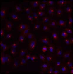
- Immunofluorescent analysis of Trans Golgi Network 46 using anti-Trans Golgi Network 46 polyclonal antibody (Product # PA1-1069) shows staining in A549 Cells.

- Submitted by
- Invitrogen Antibodies (provider)
- Main image
- Experimental details
- Immunofluorescent analysis of Trans Golgi Network 46 using anti-Trans Golgi Network 46 polyclonal antibody (Product # PA1-1069) shows staining in HMVEC Cells.

- Submitted by
- Invitrogen Antibodies (provider)
- Main image
- Experimental details
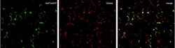
- Immunofluorescence analysis of TGN46 in MCF7 cells. MCF7 cells ectopically expressing GALT1-eGFP were stained with a TGN46 polyclonal antibody (Product # PA1-1069) at a dilution of 1:100, with an incubation overnight at 4C, washed extensively followed by an incubation with anti-rabbit secondary antibody at RT for one hour. Confocal imaging was performed at 60X magnification. Green: eGFP tagged GALT1; Red: TGN46 staining. Data courtesy of Dr Wei Xu's lab at University of Wisconsin Madison.
