Explore
 Validate
 Learn
Learn Western blot
 Immunocytochemistry
Antibody data
- Antibody Data
- Antigen structure
- References [1]
- Comments [0]
- Validations
- Immunocytochemistry [5]
- Immunohistochemistry [1]
- Other assay [1]
Submit
Validation data
Reference
Comment
Report error
- Product number
- PA5-28240 - Provider product page

- Provider
- Invitrogen Antibodies
- Product name
- SGLT1 Polyclonal Antibody
- Antibody type
- Polyclonal
- Antigen
- Recombinant full-length protein
- Description
- Recommended positive controls: 293T, A431, Jurkat, Raji. Predicted reactivity: Mouse (91%), Rat (91%), Zebrafish (80%), Dog (90%), Cat (89%), Pig (88%), Rabbit (90%), Chicken (82%), Sheep (90%), Bovine (89%). Store product as a concentrated solution. Centrifuge briefly prior to opening the vial.
- Reactivity
- Human, Mouse
- Host
- Rabbit
- Isotype
- IgG
- Vial size
- 100 μL
- Concentration
- 0.26 mg/mL
- Storage
- Store at 4°C short term. For long term storage, store at -20°C, avoiding freeze/thaw cycles.
Submitted references Empagliflozin Ammeliorates High Glucose Induced-Cardiac Dysfuntion in Human iPSC-Derived Cardiomyocytes.
Ng KM, Lau YM, Dhandhania V, Cai ZJ, Lee YK, Lai WH, Tse HF, Siu CW
Scientific reports 2018 Oct 5;8(1):14872
Scientific reports 2018 Oct 5;8(1):14872
No comments: Submit comment
Supportive validation
- Submitted by
- Invitrogen Antibodies (provider)
- Main image
- Experimental details
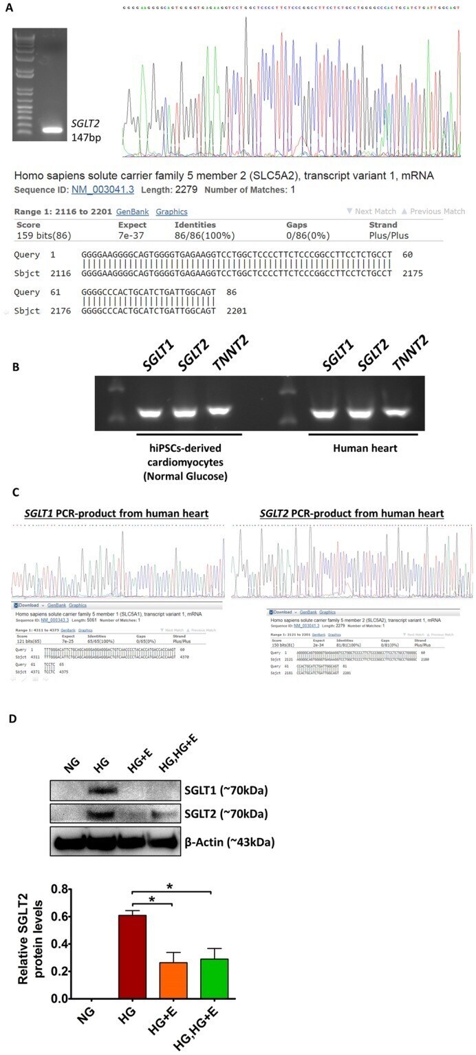
- Immunofluorescent analysis of SGLT1 in methanol-fixed A431 cells using a SGLT1 polyclonal antibody (Product # PA5-28240) at a 1:500 dilution.
- Submitted by
- Invitrogen Antibodies (provider)
- Main image

- Experimental details
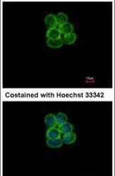
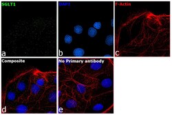
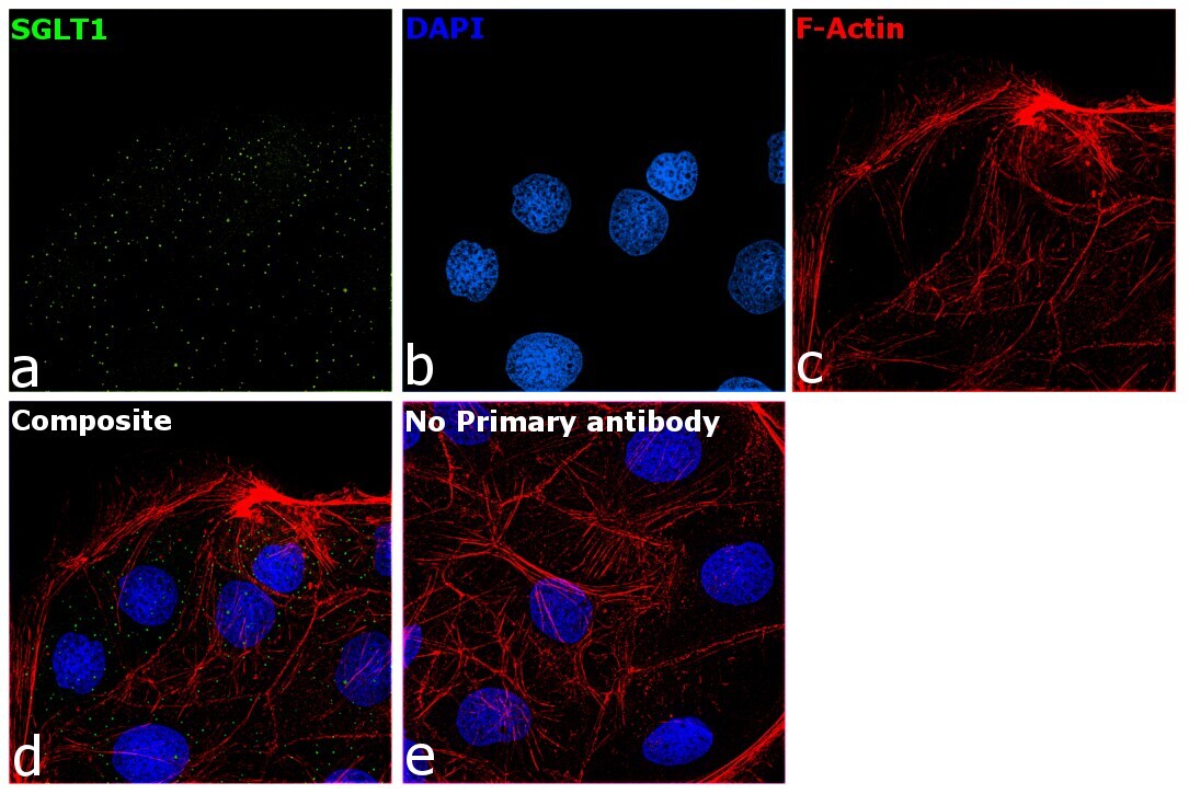
- Immunofluorescence analysis of Sodium/glucose cotransporter 1 was performed using 70% confluent log phase Caco-2 cells. The cells were fixed with 4% paraformaldehyde for 5 minutes, permeabilized with 0.1% Triton™ X-100 for 10 minutes, and blocked with 2% BSA for 45 minutes at room temperature. The cells were labeled with SGLT1 Polyclonal Antibody (Product # PA5-28240) at 1:100 dilution in 0.1% BSA, incubated at 4 degree celsius overnight and then labeled with Donkey anti-Rabbit IgG (H+L) Highly Cross-Adsorbed Secondary Antibody, Alexa Fluor Plus 488 (Product # A32790), (1:2000 dilution), for 45 minutes at room temperature (Panel a: Green). Nuclei (Panel b:Blue) were stained with ProLong™ Diamond Antifade Mountant with DAPI (Product # P36962). F-actin (Panel c: Red) was stained with Rhodamine Phalloidin (Product # R415, 1:300). Panel d represents the merged image showing punctate vesicular localization. Panel e represents control cells with no primary antibody to assess background. The images were captured at 60X magnification.

- Submitted by
- Invitrogen Antibodies (provider)
- Main image
- Experimental details
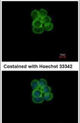
- Knockdown of Sodium/glucose cotransporter 1 was achieved by transfecting Caco-2 cells with Sodium/glucose cotransporter 1 specific siRNA (Silencer® select Product # S12952, S12953). Immunofluorescence analysis was performed on untransfected Caco-2 cells (panel a,d), transfected with non-specific scrambled siRNA (panels b,e) and transfected with Sodium/glucose cotransporter 1 specific siRNA (panel c,f). Cells were fixed, permeabilized, and labelled with SGLT1 Polyclonal Antibody (Product # PA5-28240, 1:100 dilution) followed by Donkey anti-Rabbit IgG (H+L) Highly Cross-Adsorbed Secondary Antibody, Alexa Fluor Plus 488 (Product # A32790), (1:2000 dilution). Nuclei (blue) were stained using ProLong™ Diamond Antifade Mountant with DAPI (Product # P36962), and Rhodamine Phalloidin (Product # R415, 1:300) was used for cytoskeletal F-actin (Red) staining. Decrease in the vesicular punctate specific signal was observed upon siRNA mediated knockdown (panel c,f) confirming specificity of the antibody to Sodium/glucose cotransporter 1 (Green). The Images were captured at 60X magnification.

- Submitted by
- Invitrogen Antibodies (provider)
- Main image

- Experimental details
- Knockdown of Sodium/glucose cotransporter 1 was achieved by transfecting Caco-2 cells with Sodium/glucose cotransporter 1 specific siRNA (Silencer® select Product # S12952, S12953). Immunofluorescence analysis was performed on untransfected Caco-2 cells (panel a,d), transfected with non-specific scrambled siRNA (panels b,e) and transfected with Sodium/glucose cotransporter 1 specific siRNA (panel c,f). Cells were fixed, permeabilized, and labelled with SGLT1 Polyclonal Antibody (Product # PA5-28240, 1:100 dilution) followed by Donkey anti-Rabbit IgG (H+L) Highly Cross-Adsorbed Secondary Antibody, Alexa Fluor Plus 488 (Product # A32790), (1:2000 dilution). Nuclei (blue) were stained using ProLong™ Diamond Antifade Mountant with DAPI (Product # P36962), and Rhodamine Phalloidin (Product # R415, 1:300) was used for cytoskeletal F-actin (Red) staining. Decrease in the vesicular punctate specific signal was observed upon siRNA mediated knockdown (panel c,f) confirming specificity of the antibody to Sodium/glucose cotransporter 1 (Green). The Images were captured at 60X magnification.

- Submitted by
- Invitrogen Antibodies (provider)
- Main image
- Experimental details
- Immunofluorescence analysis of Sodium/glucose cotransporter 1 was performed using 70% confluent log phase Caco-2 cells. The cells were fixed with 4% paraformaldehyde for 5 minutes, permeabilized with 0.1% Triton™ X-100 for 10 minutes, and blocked with 2% BSA for 45 minutes at room temperature. The cells were labeled with SGLT1 Polyclonal Antibody (Product # PA5-28240) at 1:100 dilution in 0.1% BSA, incubated at 4 degree celsius overnight and then labeled with Donkey anti-Rabbit IgG (H+L) Highly Cross-Adsorbed Secondary Antibody, Alexa Fluor Plus 488 (Product # A32790), (1:2000 dilution), for 45 minutes at room temperature (Panel a: Green). Nuclei (Panel b:Blue) were stained with ProLong™ Diamond Antifade Mountant with DAPI (Product # P36962). F-actin (Panel c: Red) was stained with Rhodamine Phalloidin (Product # R415, 1:300). Panel d represents the merged image showing punctate vesicular localization. Panel e represents control cells with no primary antibody to assess background. The images were captured at 60X magnification.
Supportive validation
- Submitted by
- Invitrogen Antibodies (provider)
- Main image
- Experimental details
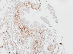
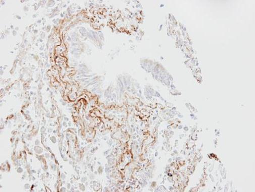
- Immunohistochemical analysis of paraffin-embedded human lung fibrose tissue, using SGLT1 (Product # PA5-28240) antibody at 1:100 dilution. Antigen Retrieval: EDTA based buffer, pH 8.0, 15 min.
Supportive validation
- Submitted by
- Invitrogen Antibodies (provider)
- Main image
- Experimental details
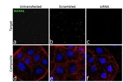
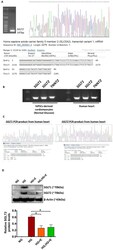
- Figure 6 Expression of SGLT1 and SGLT2 in hiPSC-derived cardiomyocytes and human heart tissue. ( A ) Confirmation of the identity of the PCR product obtained from SGLT2 -specific amplifications by DNA sequencing analysis. (B , C) Expression of SGLT1 and SGLT2 in human heart tissue were confirmed by PCR analysis and DNA sequencing analysis. (D) Protein levels of SGLT1 and SGLT2 in the hiPSC-derived cardiomyocytes were evaluated by Western blot analysis. *p < 0 . 05 . For the full-length images of gels and blots shown in this figure, please refer to Supplementary Figs S4 and S5 .