Explore
 Validate
 Learn
Learn Western blot
 Flow cytometry
Antibody data
- Antibody Data
- Antigen structure
- References [1]
- Comments [0]
- Validations
- Flow cytometry [1]
- Other assay [2]
Submit
Validation data
Reference
Comment
Report error
- Product number
- PA5-80231 - Provider product page

- Provider
- Invitrogen Antibodies
- Product name
- WNT7A Polyclonal Antibody
- Antibody type
- Polyclonal
- Antigen
- Synthetic peptide
- Description
- Reconstitute with 0.2 mL of distilled water to yield a concentration of 500 µg/mL. Positive Control - WB: human hepatocellular carcinoma tumor tissue (HCCT), human hepatocellular carcinoma paracancerous tissue (HCCP)rat kidney tissue, rat liver tissue, mouse kidney tissue, mouse liver tissue. Flow: PC-3 cell.
- Reactivity
- Human, Mouse, Rat
- Host
- Rabbit
- Isotype
- IgG
- Vial size
- 100 μg
- Concentration
- 500 μg/mL
- Storage
- -20°C
Submitted references Selective Surface and Intraluminal Localization of Wnt Ligands on Small Extracellular Vesicles Released by HT-22 Hippocampal Neurons.
Torres VI, Barrera DP, Varas-Godoy M, Arancibia D, Inestrosa NC
Frontiers in cell and developmental biology 2021;9:735888
Frontiers in cell and developmental biology 2021;9:735888
No comments: Submit comment
Supportive validation
- Submitted by
- Invitrogen Antibodies (provider)
- Main image

- Experimental details
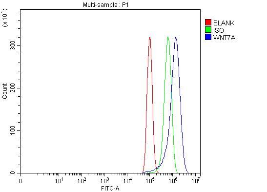
- Flow cytometry analysis of WNT7A in PC-3 cells using WNT7A Polyclonal Antibody (Product # PA5-80231), shown in overlay histogram (blue line). The cells were fixed with 4% paraformaldehyde and blocked with 10% normal goat serum, and incubated with the primary antibody (1 μg/1x10^6 cells) for 30 min at 20°C. DyLight 488 conjugated goat anti-rabbit IgG (5-10 µg/1x10^6 cells) was used as secondary antibody for 30 minutes at 20°C. Isotype control antibody (Green line) was rabbit IgG (1 µg/1x10^6) used under the same conditions. Unlabelled sample without incubation with primary antibody and secondary antibody (Red line) was used as a blank control.
Supportive validation
- Submitted by
- Invitrogen Antibodies (provider)
- Main image
- Experimental details
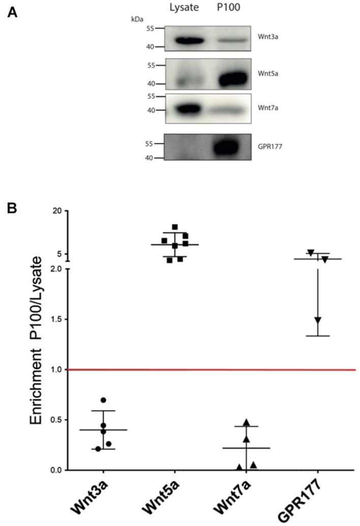
- FIGURE 2 Wnt ligand expression levels in HT-22 cells. (A) Comparison of western blot analysis of Wnt3a, Wnt5a, Wnt7a, and GPR177 protein content in cell lysate and P100 fraction. Each lane was loaded with 30 mug protein. (B) Summary densitometric measurement of band intensity showing enrichment (fold) of each protein in the P100 pellet relative to a corresponding lysate. Wnt3a, 0.4 +- 0.19 ( n = 5); Wnt5a, 8.70 +- 4.14 ( n = 7); Wnt7a, 0.22 +- 0.21 ( n = 4; four independent cell culture preparations); GPR177 3.30 +- 1.93 ( n = 3; independent cell culture preparations).
- Submitted by
- Invitrogen Antibodies (provider)
- Main image
- Experimental details
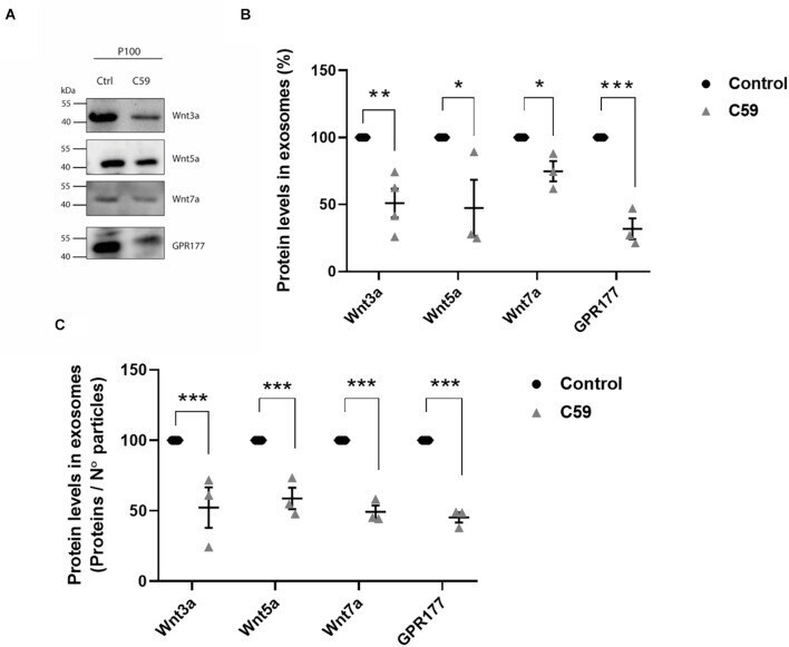
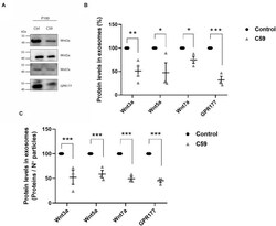
- FIGURE 5 Effects of Wnt-C59 on the content of Wnt ligands in the P100 fraction. (A) Comparison of the western blot analysis of Wnt3a, Wnt5a, Wnt7a, and GPR177 protein expression levels in the P100 fraction derived from control and Wnt-C59 treated cells (each lane was loaded with 30 mug protein). (B) Relative protein expression levels of Wnt ligands and GPR177 in the P100 fraction. The staining intensity of each band derived from control P100 was assigned a value of 100%, and the value obtained in the P100 from Wnt-C59 treatment was compared with its respective control. Wnt3a (Control: 100%, Wnt-C59: 51.1 +- 21.5%; n = 3; p = 0.0039); Wnt5a (Control: 100%, Wnt-C59: 39.1 +- 45.6%; n = 3; p = 0.0404); Wnt7a (Control: 100%, Wnt-C59: 74.82 +- 13.1%; n = 3; p = 0.0291); GPR177 (Control: 100%, Wnt-C59: 31.9 +- 13.6%; n = 3; p = 0.001). (C) Protein expression levels of Wnt ligands and GPR177 relative to the number of particles loaded per well. Control P100 was assigned a value of 100%, and the value obtained in the P100 from Wnt-C59 treatment was compared with its respective control. In both types of analysis of (B,C) , the Wnt3a, Wnt5a, Wnt7a, and GPR177 contents significantly decreased in the exosomes secreted by Wnt-C59 treated cells relative to their levels in the control cells based on the results of Mann-Whitney non-parametric t -student test (ns, not significant; * p < 0.05; ** p < 0.01; *** p < 0.005). Wnt3a (Control: 100%, Wnt-C59: 52.4 +- 24.9%; n = 3; p = 0.0295); Wnt5a (C