 Explore
 Validate
 Learn
Learn Western blot
Antibody data
- Antibody Data
- Antigen structure
- References [0]
- Comments [0]
- Validations
- Western blot [4]
- Immunocytochemistry [3]
Submit
Validation data
Reference
Comment
Report error
- Product number
- PA5-47617 - Provider product page

- Provider
- Invitrogen Antibodies
- Product name
- ID1 Polyclonal Antibody
- Antibody type
- Polyclonal
- Antigen
- Recombinant full-length protein
- Description
- Reconstitute at 0.2 mg/mL in sterile PBS.
- Reactivity
- Human, Mouse
- Host
- Goat
- Isotype
- IgG
- Vial size
- 100 µg
- Concentration
- 0.2 mg/mL
- Storage
- -20° C, Avoid Freeze/Thaw Cycles
No comments: Submit comment
Supportive validation
- Submitted by
- Invitrogen Antibodies (provider)
- Main image
- Experimental details
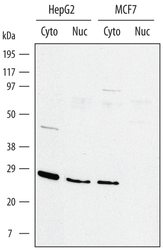
- Western blot analysis from lysates of HepG2 human hepatocellular carcinoma cell line and MCF-7 human breast cancer cell line. Gels were loaded with 20 µg of cytoplasmic (Cyto) and 10 µg of nuclear extracts (Nuc). PVDF membrane was probed with 1 µg/mL Goat Anti-human/mouse ID1 Antigen Affinity-purified Polyclonal Antibody (Product # PA5-47617) followed by HRP-conjugated Anti-Goat IgG Secondary Antibody. A specific band for ID1 was detected at approximately 25 kDa (as indicated). This experiment was conducted under reducing conditions.

- Submitted by
- Invitrogen Antibodies (provider)
- Main image
- Experimental details
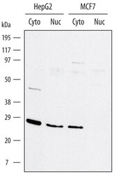
- Western blot analysis of ID1 in HepG2 human hepatocellular carcinoma cell line and MCF-7 human breast cancer cell line. Samples were incubated in ID1 polyclonal antibody (Product # PA5-47617) using a dilution of 1 µg/mL followed by a HRP-conjugated Anti-Goat IgG secondary antibody. Gels were loaded with 20 μg of cytoplasmic (Cyto) and 10 μg of nuclear extracts (Nuc). A specific band for ID1 was detected at approximately 25 kDa (as indicated). This experiment was conducted under reducing conditions.

- Submitted by
- Invitrogen Antibodies (provider)
- Main image
- Experimental details
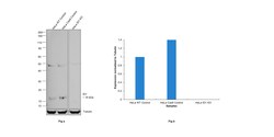
- Knockout of ID1 was achieved by CRISPR-Cas9 genome editing using LentiArray™ Lentiviral sgRNA (Product # A32042, Assay ID CRISPR627090_LV) and LentiArray Cas9 Lentivirus (Product # A32064). Western blot analysis of ID1 was performed by loading 60 µg of HeLa wild type (Lane 1), HeLa Cas9 (Lane 2) andHeLa ID1 KO (Lane 3) whole cell extracts. The samples were electrophoresed using NuPAGE™ Novex™ 4-12% Bis-Tris Protein Gel (Product # NP0322BOX). Resolved proteins were then transferred onto a nitrocellulose membrane (Product # IB23001) by iBlot® 2 Dry Blotting System (Product # IB21001). The blot was probed with Anti-ID1 Polyclonal Antibody (Product # PA5-47617, 1 µg/mL dilution) and Rabbit anti-Goat IgG (H+L) Superclonal™ Recombinant Secondary Antibody, HRP (Product # A27014, 1:5000 dilution) using the iBright™ FL 1500 (Product # A44115). Chemiluminescent detection was performed using SuperSignal™ West Dura Extended Duration Substrate (Product # 34076). Loss of signal upon CRISPR mediated knockout (KO) using the LentiArray™ CRISPR product line confirms that antibody is specific to ID1. Uncharacterized band was observed in all the samples at ~49 kDa.

- Submitted by
- Invitrogen Antibodies (provider)
- Main image

- Experimental details
- Western blot analysis of ID1 in 0.2 mg/mL lysates of HepG2 human hepatocellular carcinoma cell line. Samples were incubated in ID1 polyclonal antibody (Product # PA5-47617) using a dilution of 20 µg/mL. A specific band was detected for ID1 at approximately 30 kDa (as indicated). This experiment was conducted under reducing conditions and using the 12-230 kDa separation system.

Supportive validation
- Submitted by
- Invitrogen Antibodies (provider)
- Main image
- Experimental details
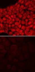
- Immunocytochemistry analysis of ID1 in immersion fixed BG01V human embryonic stem cells, undifferentiated (lower panel) and differentiated into neural progenitor cells (upper panel). Samples were incubated in ID1 polyclonal antibody (Product # PA5-47617) using a dilution of 10 µg/mL for 3 hours at room temperature followed by NorthernLights™ 557-conjugated Anti-Goat IgG Secondary Antibody (red) and counterstained with DAPI (blue). Specific staining was localized to nuclei.

- Submitted by
- Invitrogen Antibodies (provider)
- Main image

- Experimental details
- Immunofluorescence analysis of ID1 was performed using 70% confluent log phase MCF7 cells. The cells were fixed with 4% paraformaldehyde for 10 minutes, permeabilized with 0.1% Triton™ X-100 for 15 minutes, and blocked with 2% BSA for 45 minutes at room temperature. The cells were labeled with ID1 Polyclonal Antibody (Product # PA5-47617) at 10 µg/mL dilution in 0.1% BSA, incubated at 4 degree celsius overnight and then labeled with Donkey anti-Goat IgG (H+L) Highly Cross-Adsorbed Secondary Antibody, Alexa Fluor Plus 488 (Product # A32814), (1:2000 dilution), for 45 minutes at room temperature (Panel a: Green). Nuclei (Panel b: Blue) were stained with ProLong™ Diamond Antifade Mountant with DAPI (Product # P36962). F-actin (Panel c: Red) was stained with Rhodamine Phalloidin (Product # R415, 1:300). Panel d represents the merged image showing predominantly nuclear and faint cytoplasmic localization. Panel e represents U-937 cells with no expression of ID1. Panel f represents control cells with no primary antibody to assess background. The images were captured at 60X.
