Explore
 Validate
 Learn
Learn Western blot
Antibody data
- Antibody Data
- Antigen structure
- References [2]
- Comments [0]
- Validations
- Western blot [1]
- Immunocytochemistry [1]
Submit
Validation data
Reference
Comment
Report error
- Product number
- AF4377 - Provider product page

- Provider
- R&D Systems
- Product name
- Human/Mouse ID1 Antibody
- Antibody type
- Polyclonal
- Description
- Immunogen affinity purified. Detects human and mouse ID1 in Western blots.
- Reactivity
- Human, Mouse
- Host
- Goat
- Conjugate
- Unconjugated
- Antigen sequence
P20067- Isotype
- IgG
- Vial size
- 100 ug
- Concentration
- LYOPH
- Storage
- Use a manual defrost freezer and avoid repeated freeze-thaw cycles. 12 months from date of receipt, -20 to -70 °C as supplied. 1 month, 2 to 8 °C under sterile conditions after reconstitution. 6 months, -20 to -70 °C under sterile conditions after reconstitution.
Submitted references Suppression of ID1 expression in colon cancer cells increases sensitivity to 5-fluorouracil.
Characterization of BMS-911543, a functionally selective small-molecule inhibitor of JAK2.
Przybyła T, Sakowicz-Burkiewicz M, Maciejewska I, Bielarczyk H, Pawełczyk T
Acta biochimica Polonica 2017;64(2):315-322
Acta biochimica Polonica 2017;64(2):315-322
Characterization of BMS-911543, a functionally selective small-molecule inhibitor of JAK2.
Purandare AV, McDevitt TM, Wan H, You D, Penhallow B, Han X, Vuppugalla R, Zhang Y, Ruepp SU, Trainor GL, Lombardo L, Pedicord D, Gottardis MM, Ross-Macdonald P, de Silva H, Hosbach J, Emanuel SL, Blat Y, Fitzpatrick E, Taylor TL, McIntyre KW, Michaud E, Mulligan C, Lee FY, Woolfson A, Lasho TL, Pardanani A, Tefferi A, Lorenzi MV
Leukemia 2012 Feb;26(2):280-8
Leukemia 2012 Feb;26(2):280-8
No comments: Submit comment
Supportive validation
- Submitted by
- R&D Systems (provider)
- Main image
- Experimental details
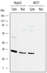
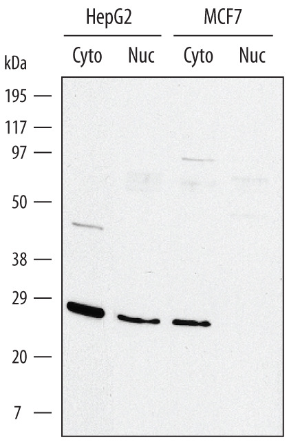
- Detection of Human ID1 by Western Blot. Western blot shows lysates of HepG2 human hepatocellular carcinoma cell line and MCF-7 human breast cancer cell line. Gels were loaded with 20 μg of cytoplasmic (Cyto) and 10 μg of nuclear extracts (Nuc). PVDF membrane was probed with 1 µg/mL Goat Anti-Human/Mouse ID1 Antigen Affinity-purified Polyclonal Antibody (Catalog # AF4377) followed by HRP-conjugated Anti-Goat IgG Secondary Antibody (Catalog # HAF017). A specific band for ID1 was detected at approximately 25 kDa (as indicated). This experiment was conducted under reducing conditions and using Immunoblot Buffer Group 1.
Supportive validation
- Submitted by
- R&D Systems (provider)
- Main image
- Experimental details
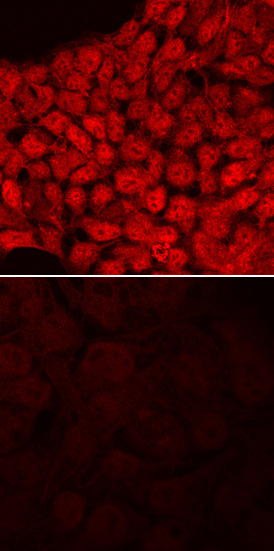
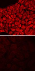
- ID1 in BG01V Human Embryonic Stem Cells. ID1 was detected in immersion fixed BG01V human embryonic stem cells, undifferentiated (lower panel) and differentiated into neural progenitor cells (upper panel), using Goat Anti-Human/Mouse ID1 Antigen Affinity-purified Polyclonal Antibody (Catalog # AF4377) at 10 µg/mL for 3 hours at room temperature. Cells were stained using the NorthernLights™ 557-conjugated Anti-Goat IgG Secondary Antibody (red; Catalog # NL001) and counterstained with DAPI (blue). Specific staining was localized to nuclei. View our protocol for Fluorescent ICC Staining of Cells on Coverslips.