Explore
 Validate
 Learn
Learn Western blot
 ELISA
ELISAAntibody data
- Antibody Data
- Antigen structure
- References [1]
- Comments [0]
- Validations
- Western blot [1]
- Immunohistochemistry [1]
- Other assay [1]
Submit
Validation data
Reference
Comment
Report error
- Product number
- PA5-98885 - Provider product page

- Provider
- Invitrogen Antibodies
- Product name
- ARL8B Polyclonal Antibody
- Antibody type
- Polyclonal
- Antigen
- Recombinant full-length protein
- Reactivity
- Human, Mouse
- Host
- Rabbit
- Isotype
- IgG
- Vial size
- 100 μg
- Concentration
- 1 mg/mL
- Storage
- -20°C or -80°C if preferred
Submitted references The amyloid plaque proteome in early onset Alzheimer's disease and Down syndrome.
Drummond E, Kavanagh T, Pires G, Marta-Ariza M, Kanshin E, Nayak S, Faustin A, Berdah V, Ueberheide B, Wisniewski T
Acta neuropathologica communications 2022 Apr 13;10(1):53
Acta neuropathologica communications 2022 Apr 13;10(1):53
No comments: Submit comment
Supportive validation
- Submitted by
- Invitrogen Antibodies (provider)
- Main image

- Experimental details
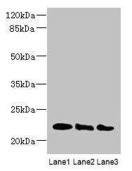
- Western Blot analysis of ARL8B using a ARL8B Polyclonal antibody (Product # PA5-98885) at a concentration of 4 µg/mL. Lane 1: Mouse brain tissue. Lane 2: NIH/3T3 whole cell lysate. Lane 3: Jurkat whole cell lysate. A secondary Goat polyclonal antibody to rabbit IgG was applied at a 1:10,000 dilution. Observed band size: 22 kDa.
Supportive validation
- Submitted by
- Invitrogen Antibodies (provider)
- Main image
- Experimental details
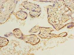
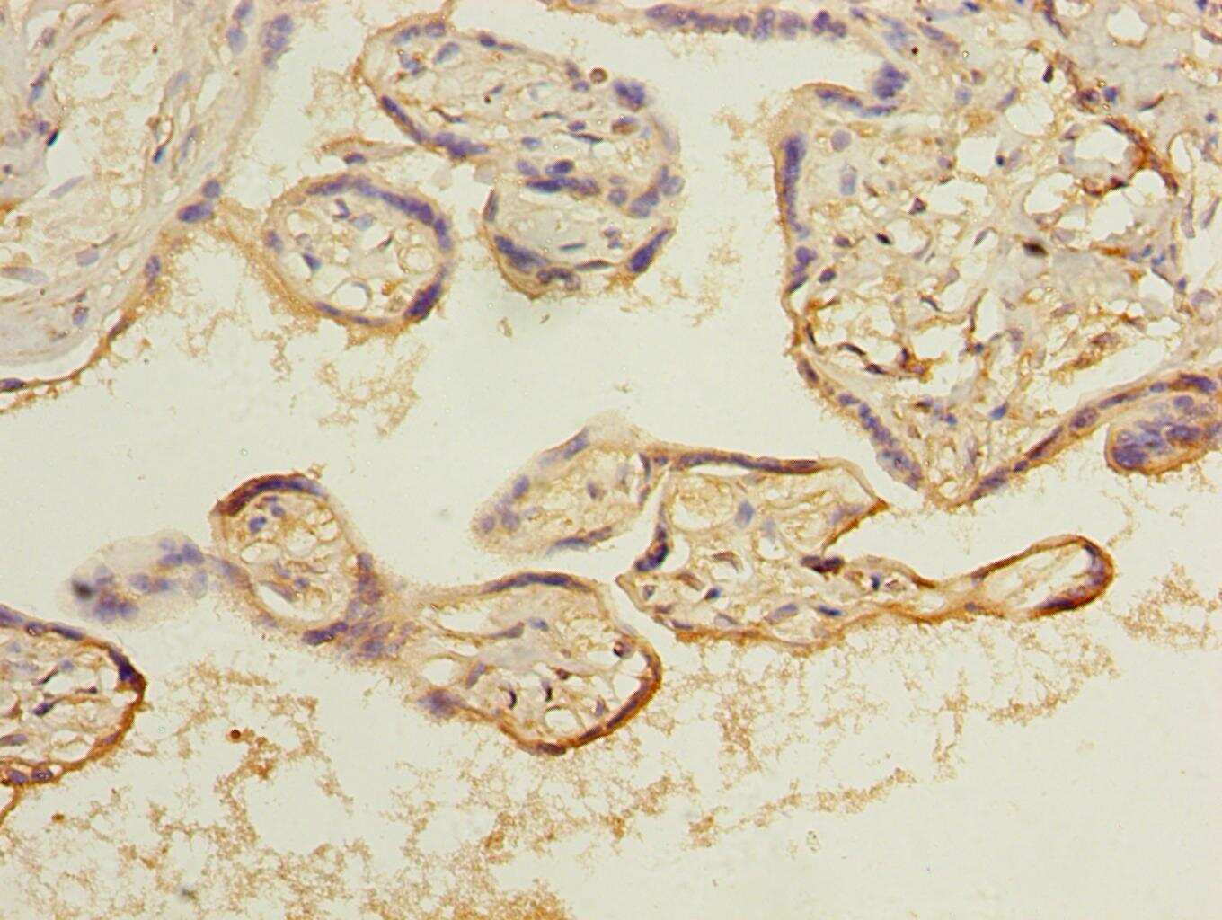
- Immunohistochemical analysis of ARL8B in paraffin embedded human placenta tissue using a ARL8B polyclonal antibody (Product # PA5-98885) at a dilution of 1:100.
Supportive validation
- Submitted by
- Invitrogen Antibodies (provider)
- Main image

- Experimental details
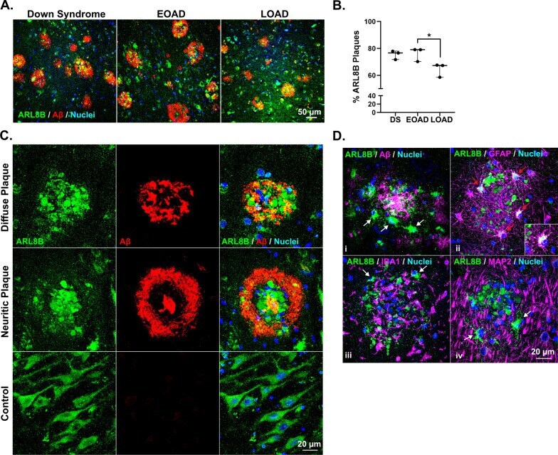
- Validation of ARL8B as a plaque enriched protein in human brain tissue by immunohistochemistry. A Enrichment of ARL8B in amyloid plaques was observed in DS, EOAD and LOAD cases. B Plot shows percentage of ARL8B immunoreactive plaques in the hippocampus of DS, EOAD and LOAD cases (n = 3/group). Results generated by an analysis of 309 +- 41 hippocampal plaques (average +- SEM) in each case. The ratio of ARL8B positive plaques (immunoreactive for both Abeta and ARL8B) over the total number of amyloid plaques was calculated for each case in DS, EOAD and LOAD. C Representative images showing ARL8B distribution in amyloid plaques. Bright puncta of ARL8B were diffusely present throughout both diffuse and neuritic plaques. Basal ARL8B staining was observed in controls in neuron soma. D Intense ARL8B immunoreactivity was observed in plaque-associated cells (i; arrows). Double-fluorescent immunohistochemistry showed that these plaque-associated cells with intense ARL8B immunoreactivity were a subset of plaque-associated reactive astrocytes (ii; GFAP, red arrows), and not plaque associated reactive microglia (iii; IBA1, white arrows) or neurons (iv; MAP2, white arrows). Insert in ii shows a higher magnification image of the colocalization of ARL8B and GFAP in plaque associated astrocytes