Explore
 Validate
 Learn
Learn Western blot
 Immunocytochemistry
Antibody data
- Antibody Data
- Antigen structure
- References [2]
- Comments [0]
- Validations
- Immunocytochemistry [1]
- Immunohistochemistry [1]
Submit
Validation data
Reference
Comment
Report error
- Product number
- HPA029102 - Provider product page

- Provider
- Atlas Antibodies
- Proper citation
- Atlas Antibodies Cat#HPA029102, RRID:AB_10599846
- Product name
- Anti-RGN
- Antibody type
- Polyclonal
- Description
- Polyclonal Antibody against Human RGN, Gene description: regucalcin, Alternative Gene Names: RC, SMP30, Validated applications: WB, IHC, ICC, Uniprot ID: Q15493, Storage: Store at +4°C for short term storage. Long time storage is recommended at -20°C.
- Reactivity
- Human
- Host
- Rabbit
- Conjugate
- Unconjugated
- Isotype
- IgG
- Vial size
- 100 µl
- Concentration
- 0.1 mg/ml
- Storage
- Store at +4°C for short term storage. Long time storage is recommended at -20°C.
- Handling
- The antibody solution should be gently mixed before use.
Submitted references The phytochemical p-hydroxycinnamic acid suppresses the growth and stimulates the death in human liver cancer HepG2 cells
Affinity Proteomics Exploration of Melanoma Identifies Proteins in Serum with Associations to T-Stage and Recurrence.
Yamaguchi M, Murata T, Ramos J
Anti-Cancer Drugs 2021;32(5):558-566
Anti-Cancer Drugs 2021;32(5):558-566
Affinity Proteomics Exploration of Melanoma Identifies Proteins in Serum with Associations to T-Stage and Recurrence.
Byström S, Fredolini C, Edqvist PH, Nyaiesh EN, Drobin K, Uhlén M, Bergqvist M, Pontén F, Schwenk JM
Translational oncology 2017 Jun;10(3):385-395
Translational oncology 2017 Jun;10(3):385-395
No comments: Submit comment
Supportive validation
- Submitted by
- Atlas Antibodies (provider)
- Main image
- Experimental details
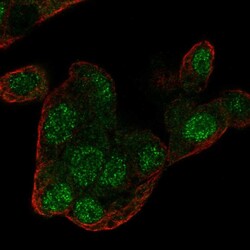
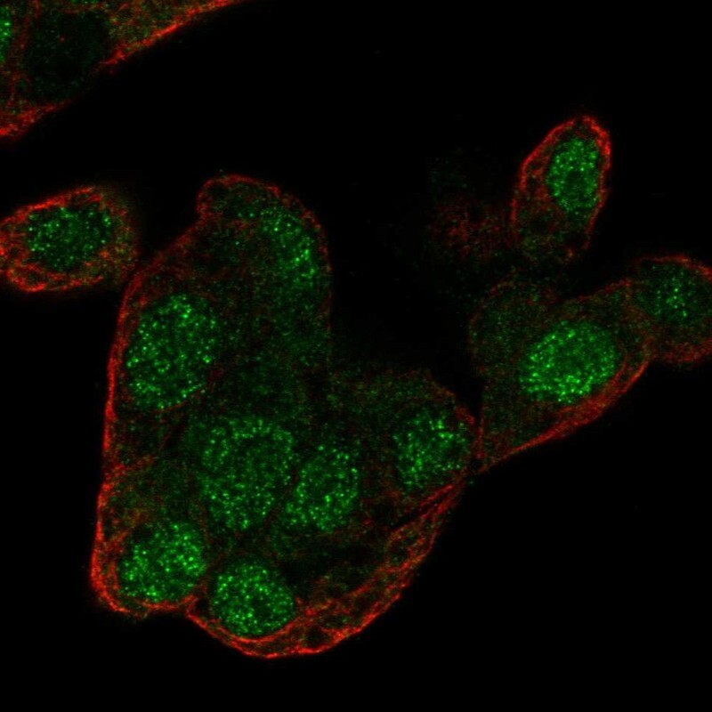
- Immunofluorescent staining of human cell line Hep G2 shows localization to nucleus.
- Sample type
- Human
Supportive validation
- Submitted by
- Atlas Antibodies (provider)
- Enhanced method
- Orthogonal validation
- Main image
- Experimental details
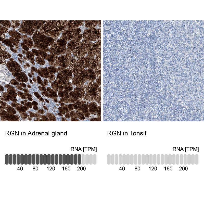
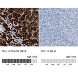
- Immunohistochemistry analysis in human adrenal gland and tonsil tissues using HPA029102 antibody. Corresponding RGN RNA-seq data are presented for the same tissues.
- Sample type
- Human
- Protocol
- Protocol