Explore
 Validate
 Learn
Learn Western blot
Antibody data
- Antibody Data
- Antigen structure
- References [0]
- Comments [0]
- Validations
- Western blot [2]
- Immunohistochemistry [1]
Submit
Validation data
Reference
Comment
Report error
- Product number
- ANR-017-25UL - Provider product page

- Provider
- Invitrogen Antibodies
- Product name
- Synapsin III (SYN3) Polyclonal Antibody
- Antibody type
- Polyclonal
- Antigen
- Other
- Reactivity
- Human, Mouse, Rat
- Host
- Rabbit
- Isotype
- IgG
- Vial size
- 25 µL
- Concentration
- 0.8 mg/mL
- Storage
- -20° C, Avoid Freeze/Thaw Cycles
No comments: Submit comment
Supportive validation
- Submitted by
- Invitrogen Antibodies (provider)
- Main image
- Experimental details
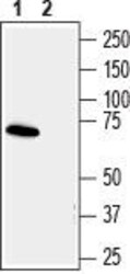
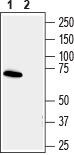
- Western blot analysis human SH-SY5Y neuroblastoma cell line lysate: - 1. Anti-Synapsins III (SYN3) Antibody (#ANR-017), (1:200). 2. Anti-Synapsins III (SYN3) Antibody , preincubated with Synapsins III/SYN3 Blocking Peptide (#BLP-NR017).
- Submitted by
- Invitrogen Antibodies (provider)
- Main image
- Experimental details
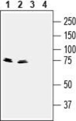
- Western blot analysis of mouse (lanes 1 and 3) and rat (lanes 2 and 4)brain lysates: - 1,2. Anti-Synapsins III (SYN3) Antibody (#ANR-017), (1:400).3,4. Anti-Synapsins III (SYN3) Antibody , preincubated with Synapsins III/SYN3 Blocking Peptide (#BLP-NR017).

Supportive validation
- Submitted by
- Invitrogen Antibodies (provider)
- Main image
- Experimental details
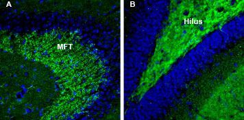
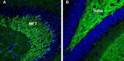
- Expression of Synapsins 3 in mouse and rat hippocampus - Immunohistochemical staining of perfusion-fixed frozen mouse brain sections with Anti-Synapsins III (SYN3) Antibody (#ANR-017), (1:200), followed by goat Anti-rabbit-AlexaFluor-488. A. SYN3 staining (green) in mouse hippocampal CA3 region is detected in the mossy fiber terminal (MFT) region. B. In rat hippocampal dentate gyrus (DG) region, SYN3 immunoreactivity (green) is observed in hilus of the dentate gyrus. Cell nuclei are stained with DAPI (blue).