Explore
 Validate
 Learn
Learn Western blot
Antibody data
- Antibody Data
- Antigen structure
- References [0]
- Comments [0]
- Validations
- Western blot [1]
- Immunohistochemistry [1]
- Blocking/Neutralizing [1]
Submit
Validation data
Reference
Comment
Report error
- Product number
- AF1143 - Provider product page

- Provider
- Novus Biologicals
- Product name
- Goat Polyclonal PD-ECGF/Thymidine Phosphorylase Antibody
- Antibody type
- Polyclonal
- Description
- Immunogen affinity purified. Detects human PD-ECGF/Thymidine Phosphorylase in direct ELISAs and Western blots.
- Reactivity
- Human
- Host
- Goat
- Conjugate
- Unconjugated
- Isotype
- IgG
- Vial size
- 100 ug
- Concentration
- LYOPH
- Storage
- Use a manual defrost freezer and avoid repeated freeze-thaw cycles. 12 months from date of receipt, -20 to -70 degreesC as supplied. 1 month, 2 to 8 degreesC under sterile conditions after reconstitution. 6 months, -20 to -70 degreesC under sterile conditions after reconstitution.
No comments: Submit comment
Supportive validation
- Submitted by
- Novus Biologicals (provider)
- Main image
- Experimental details
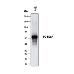
- Detection of Human PD-ECGF/Thymidine Phosphorylase by Western Blot. Detection of Human PD-ECGF/Thymidine Phosphorylase by Western Blot.

Supportive validation
- Submitted by
- Novus Biologicals (provider)
- Main image
- Experimental details
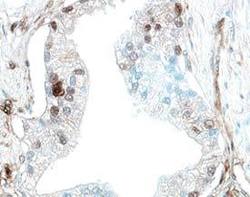
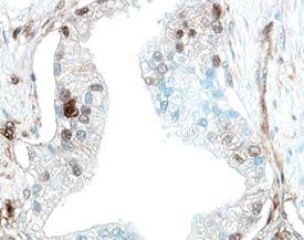
- PD-ECGF/Thymidine Phosphorylase in Human Prostate Cancer Tissue. PD-ECGF/Thymidine Phosphorylase was detected in immersion fixed paraffin-embedded sections of human prostate cancer tissue using 5 µg/mL Goat Anti-Human PD-ECGF/Thymidine Phosphorylase Antigen Affinity-purified Polyclonal Antibody (Catalog # AF1143) overnight at 4 °C. Tissue was stained with the Anti-Goat HRP-DAB Cell & Tissue Staining Kit (brown; Catalog # CTS008) and counter-stained with hematoxylin (blue). View our protocol for Chromogenic IHC Staining of Paraffin-embedded Tissue Sections.
Supportive validation
- Submitted by
- Novus Biologicals (provider)
- Main image
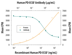
- Experimental details
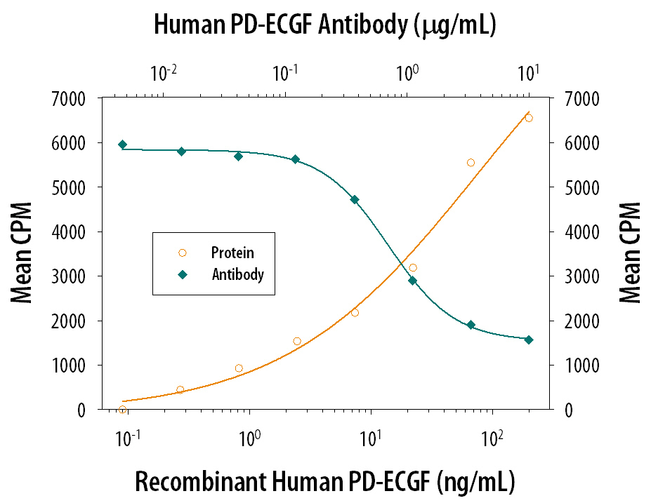
- Cell Proliferation Induced by PD-ECGF/Thymidine Phosphorylase and Neutralization by Human PD-ECGF/Thymidine Phosphorylase Antibody. Recombinant Human PD-ECGF/Thymidine Phosphorylase (Catalog # 229-PE) stimulates proliferation in HUVEC human umbilical vein endothelial cells in a dose-dependent manner (orange line). Proliferation elicited by Recombinant Human PD-ECGF/Thymidine Phosphorylase (150 ng/mL) is neutralized (green line) by increasing concentrations of Human PD-ECGF/Thymidine Phosphorylase Antigen Affinity-purified Polyclonal Antibody (Catalog # AF1143). The ND50 is typically 0.5-2.0 µg/mL.