 Explore
 Validate
 Learn
Learn Flow cytometry
Antibody data
- Antibody Data
- Antigen structure
- References [4]
- Comments [0]
- Validations
- Flow cytometry [1]
Submit
Validation data
Reference
Comment
Report error
- Product number
- 12-0019-42 - Provider product page

- Provider
- Invitrogen Antibodies
- Product name
- CD1a Monoclonal Antibody (HI149), PE, eBioscience™
- Antibody type
- Monoclonal
- Antigen
- Other
- Description
- Description: The HI149 monoclonal antibody reacts with human CD1a, a 49 kDa protein expressed by cortical thymocytes and dendritic cells including Langerhans cells. The CD1 family of proteins share some structural and functional characteristics with the MHC class I molecules; however, members of the CD1 family are not polymorphic. Similar to MHC class I, CD1a associates with the beta2-microglobulin and is thought to play a role in antigen presentation. Applications Reported: The HI149 antibody has been reported for use in flow cytometric analysis. Applications Tested: This HI149 antibody has been pre-titrated and tested by flow cytometric analysis of MOLT-4 cells. This can be used at 5 µL (0.5 µg) per test. A test is defined as the amount (µg) of antibody that will stain a cell sample in a final volume of 100 µL. Cell number should be determined empirically but can range from 10^5 to 10^8 cells/test. Excitation: 488-561 nm; Emission: 578 nm; Laser: Blue Laser, Green Laser, Yellow-Green Laser. Filtration: 0.2 µm post-manufacturing filtered.
- Reactivity
- Human
- Host
- Mouse
- Conjugate
- Yellow dye
- Isotype
- IgG
- Antibody clone number
- HI149
- Vial size
- 100 Tests
- Concentration
- 5 µL/Test
- Storage
- 4° C, store in dark, DO NOT FREEZE!
Submitted references In vitro OP9-DL1 co-culture and subsequent maturation in the presence of IL-21 generates tumor antigen-specific T cells with a favorable less-differentiated phenotype and enhanced functionality.
Regulatory Dendritic Cells Restrain NK Cell IFN-γ Production through Mechanisms Involving NKp46, IL-10, and MHC Class I-Specific Inhibitory Receptors.
Dendritic Cells (DC) Facilitate Detachment of Squamous Carcinoma Cells (SCC), While SCC Promote an Immature CD16(+) DC Phenotype and Control DC Migration.
Immunological aspects of REIC/Dkk-3 in monocyte differentiation and tumor regression.
Bonte S, de Munter S, Billiet L, Goetgeluk G, Ingels J, Jansen H, Pille M, de Cock L, Weening K, Taghon T, Leclercq G, Vandekerckhove B, Kerre T
Oncoimmunology 2021;10(1):1954800
Oncoimmunology 2021;10(1):1954800
Regulatory Dendritic Cells Restrain NK Cell IFN-γ Production through Mechanisms Involving NKp46, IL-10, and MHC Class I-Specific Inhibitory Receptors.
Spallanzani RG, Torres NI, Avila DE, Ziblat A, Iraolagoitia XL, Rossi LE, Domaica CI, Fuertes MB, Rabinovich GA, Zwirner NW
Journal of immunology (Baltimore, Md. : 1950) 2015 Sep 1;195(5):2141-8
Journal of immunology (Baltimore, Md. : 1950) 2015 Sep 1;195(5):2141-8
Dendritic Cells (DC) Facilitate Detachment of Squamous Carcinoma Cells (SCC), While SCC Promote an Immature CD16(+) DC Phenotype and Control DC Migration.
Ramanathapuram LV, Hopkin D, Kurago ZB
Cancer microenvironment : official journal of the International Cancer Microenvironment Society 2013 Apr;6(1):41-55
Cancer microenvironment : official journal of the International Cancer Microenvironment Society 2013 Apr;6(1):41-55
Immunological aspects of REIC/Dkk-3 in monocyte differentiation and tumor regression.
Watanabe M, Kashiwakura Y, Huang P, Ochiai K, Futami J, Li SA, Takaoka M, Nasu Y, Sakaguchi M, Huh NH, Kumon H
International journal of oncology 2009 Mar;34(3):657-63
International journal of oncology 2009 Mar;34(3):657-63
No comments: Submit comment
Supportive validation
- Submitted by
- Invitrogen Antibodies (provider)
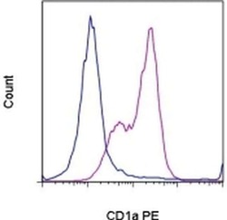
- Main image
- Experimental details
- Staining of MOLT-4 cells with Mouse IgG1 K Isotype Control PE (Product # 12-4714-81) (blue histogram) or Anti-Human CD1a PE (purple histogram). Total viable cells, as determined by Fixable Viability Dye eFluor® 660, were used for analysis.
- Conjugate
- Yellow dye
