 Explore
 Validate
 Learn
Learn Flow cytometry
Antibody data
- Antibody Data
- Antigen structure
- References [2]
- Comments [0]
- Validations
- Flow cytometry [1]
Submit
Validation data
Reference
Comment
Report error
- Product number
- 48-0019-42 - Provider product page

- Provider
- Invitrogen Antibodies
- Product name
- CD1a Monoclonal Antibody (HI149), eFluor™ 450, eBioscience™
- Antibody type
- Monoclonal
- Antigen
- Other
- Description
- Description: The HI149 monoclonal antibody reacts with human CD1a, a 49 kDa protein expressed by cortical thymocytes and dendritic cells including Langerhans cells. The CD1 family of proteins share some structural and functional characteristics with the MHC class I molecules; however, members of the CD1 family are not polymorphic. Similar to MHC class I, CD1a associates with the beta2-microglobulin and is thought to play a role in antigen presentation. Applications Reported: This HI149 antibody has been reported for use in flow cytometric analysis. Applications Tested: This HI149 antibody has been pre-titrated and tested by flow cytometric analysis of MOLT-4 cells. This can be used at 5 µL (0.5 µg) per test. A test is defined as the amount (µg) of antibody that will stain a cell sample in a final volume of 100 µL. Cell number should be determined empirically but can range from 10^5 to 10^8 cells/test. eFluor® 450 is an alternative to Pacific Blue®. eFluor® 450 emits at 445 nm and is excited with the Violet laser (405 nm). Please make sure that your instrument is capable of detecting this fluorochome. Excitation: 405 nm; Emission: 445 nm; Laser: Violet Laser. Filtration: 0.2 µm post-manufacturing filtered.
- Reactivity
- Human
- Host
- Mouse
- Isotype
- IgG
- Antibody clone number
- HI149
- Vial size
- 100 Tests
- Concentration
- 5 µL/Test
- Storage
- 4° C, store in dark, DO NOT FREEZE!
Submitted references In vitro OP9-DL1 co-culture and subsequent maturation in the presence of IL-21 generates tumor antigen-specific T cells with a favorable less-differentiated phenotype and enhanced functionality.
uPAR(+) extracellular vesicles: a robust biomarker of resistance to checkpoint inhibitor immunotherapy in metastatic melanoma patients.
Bonte S, de Munter S, Billiet L, Goetgeluk G, Ingels J, Jansen H, Pille M, de Cock L, Weening K, Taghon T, Leclercq G, Vandekerckhove B, Kerre T
Oncoimmunology 2021;10(1):1954800
Oncoimmunology 2021;10(1):1954800
uPAR(+) extracellular vesicles: a robust biomarker of resistance to checkpoint inhibitor immunotherapy in metastatic melanoma patients.
Porcelli L, Guida M, De Summa S, Di Fonte R, De Risi I, Garofoli M, Caputo M, Negri A, Strippoli S, Serratì S, Azzariti A
Journal for immunotherapy of cancer 2021 May;9(5)
Journal for immunotherapy of cancer 2021 May;9(5)
No comments: Submit comment
Supportive validation
- Submitted by
- Invitrogen Antibodies (provider)
- Main image
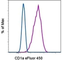
- Experimental details
- Staining of Molt-4 cells with Mouse IgG1 K Isotype Control eFluor® 450 (Product # 48-4714-82) (blue histogram) or Anti-Human CD1a eFluor® 450 (purple histogram). Total viable cells were used for analysis.
