Explore
 Validate
 Learn
Learn Immunohistochemistry
Antibody data
- Antibody Data
- Antigen structure
- References [2]
- Comments [0]
- Validations
- Immunohistochemistry [1]
Submit
Validation data
Reference
Comment
Report error
- Product number
- AMAb91081 - Provider product page

- Provider
- Atlas Antibodies
- Proper citation
- Atlas Antibodies Cat#AMAb91081, RRID:AB_2665793
- Product name
- Anti-VGLUT2
- Antibody type
- Monoclonal
- Description
- Monoclonal Antibody against Human SLC17A6, Clone ID: CL2921, Gene description: solute carrier family 17 member 6, Alternative Gene Names: SLC17A6, DNPI, Validated applications: IHC, Uniprot ID: Q9P2U8, Storage: Store at +4°C for short term storage. Long time storage is recommended at -20°C.
- Reactivity
- Human, Mouse, Rat
- Host
- Mouse
- Conjugate
- Unconjugated
- Isotype
- IgG
- Antibody clone number
- CL2921
- Vial size
- 100 µl
- Concentration
- 0.4 mg/ml
- Storage
- Store at +4°C for short term storage. Long time storage is recommended at -20°C.
- Handling
- The antibody solution should be gently mixed before use.
Submitted references Inhibition of Vesicular Glutamate Transporters (VGLUTs) with Chicago Sky Blue 6B Before Focal Cerebral Ischemia Offers Neuroprotection
Novel human pluripotent stem cell-derived hypothalamus organoids demonstrate cellular diversity.
Pomierny B, Krzyżanowska W, Skórkowska A, Jurczyk J, Bystrowska B, Budziszewska B, Pera J
Molecular Neurobiology 2023;60(6):3130-3146
Molecular Neurobiology 2023;60(6):3130-3146
Novel human pluripotent stem cell-derived hypothalamus organoids demonstrate cellular diversity.
Sarrafha L, Neavin DR, Parfitt GM, Kruglikov IA, Whitney K, Reyes R, Coccia E, Kareva T, Goldman C, Tipon R, Croft G, Crary JF, Powell JE, Blanchard J, Ahfeldt T
iScience 2023 Sep 15;26(9):107525
iScience 2023 Sep 15;26(9):107525
No comments: Submit comment
Supportive validation
- Submitted by
- Atlas Antibodies (provider)
- Enhanced method
- Orthogonal validation
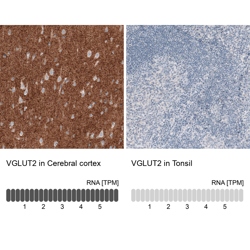
- Main image
- Experimental details
- Immunohistochemistry analysis in human cerebral cortex and tonsil tissues using AMAb91081 antibody. Corresponding VGLUT2 RNA-seq data are presented for the same tissues.
- Sample type
- Human
- Protocol
- Protocol