Explore
 Validate
 Learn
Learn Western blot
 Immunocytochemistry
 Immunohistochemistry
Antibody data
- Antibody Data
- Antigen structure
- References [1]
- Comments [0]
- Validations
- Immunocytochemistry [2]
- Other assay [1]
Submit
Validation data
Reference
Comment
Report error
- Product number
- MA5-27613 - Provider product page

- Provider
- Invitrogen Antibodies
- Product name
- VGLUT2 Monoclonal Antibody (N29/29)
- Antibody type
- Monoclonal
- Antigen
- Other
- Description
- 1 µg/mL of MA5-27613 was sufficient for detection of VGLut2 in 20 µg of rat brain lysate by colorimetric immunoblot analysis using goat anti-mouse IgG:HRP as the secondary antibody.|Detects approximately 60kDa. This antibody was formerly sold as clone S29-29
- Reactivity
- Human, Mouse, Rat
- Host
- Mouse
- Isotype
- IgG
- Antibody clone number
- N29/29
- Vial size
- 100 μg
- Concentration
- 1 mg/mL
- Storage
- -20°C
Submitted references An excitatory ventromedial hypothalamus to paraventricular thalamus circuit that suppresses food intake.
Zhang J, Chen D, Sweeney P, Yang Y
Nature communications 2020 Dec 10;11(1):6326
Nature communications 2020 Dec 10;11(1):6326
No comments: Submit comment
Supportive validation
- Submitted by
- Invitrogen Antibodies (provider)
- Main image

- Experimental details
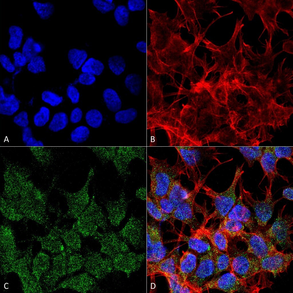
- Immunofluorescent analysis of VGLUT2 in human neuroblastoma cell line (SK-N-BE). Sample was fixed with 4% formaldehyde (15 min at RT), incubated with VGLUT2 monoclonal antibody (Product # MA5-27613) using a dilution of 1:100 (1 hour at RT), and followed by Goat Anti-Mouse 488, Phalloidin Texas Red and DAPI secondary antibody at a dilution of 1:200, 1:1000 (60 min at RT) and 1:5000 (5 min at RT). Images are shown as follows: (A) DAPI (blue) nuclear stain, B) Phalloidin Texas Red F-Actin stain, C) VGLUT2 Antibody, D) Merged image. Magnification: 60x.

- Submitted by
- Invitrogen Antibodies (provider)
- Main image

- Experimental details
- Immunofluorescent analysis of VGLUT2 in human neuroblastoma cell line (SK-N-BE). Sample was fixed with 4% formaldehyde (15 min at RT), incubated with VGLUT2 monoclonal antibody (Product # MA5-27613) using a dilution of 1:100 (1 hour at RT), and followed by Goat Anti-Mouse 488, Phalloidin Texas Red and DAPI secondary antibody at a dilution of 1:200, 1:1000 (60 min at RT) and 1:5000 (5 min at RT). Images are shown as follows: (A) DAPI (blue) nuclear stain, B) Phalloidin Texas Red F-Actin stain, C) VGLUT2 Antibody, D) Merged image. Magnification: 60x.
Supportive validation
- Submitted by
- Invitrogen Antibodies (provider)
- Main image
- Experimental details
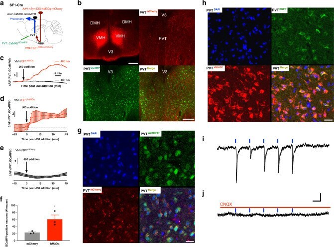
- Fig. 4 VMH SF1 neurons project to PVT neurons. a Schematic depiction of photometry monitoring of PVT neurons in SF1-Cre mice transduced with hM3Dq in VMH SF1 neurons and PVT neurons with GCaMP 6f . b - g Acute PVT brain slice photometry experiments show that DREADD activation of SF1 neurons with J60 addition in the circulating ACSF potently increased PVT GCaMP 6f signals in the SF1 neuron hM3Dq-transduced mice ( n = 4) as compared to control mCherry-transduced mice ( n = 3): b representative images of hM3Dq-transduced SF1 neurons in VMH and GCaMP 6f -positive PVT neurons; c representative real-time monitoring of GCaMP 6f signals before and after J60 addition in an isolated PVT brain slice of hM3Dq-transduced animal; d average PVT GCaMP 6f signals from the SF1 neuron hM3Dq-transduced mice ( n = 11 sections/4 mice); e average PVT neural GCaMP 6f signals from the SF1 neuron mCherry-transduced mice ( n = 9 sections/3 mice); f group data of GCaMP 6f -positive neurons in the PVT ( n = 3 per group; p = 0.0356, two-tailed Student''s t -tests); g representative confocal images of acute brain slice morphological studies show that SF1 neurons project synaptic inputs to GCaMP 6f -positive neurons in PVT, as indicated by the hM3Dq-mCherry positive SF1 terminals adjunct on GCaMP 6f -positive PVT neurons; 1 h after J60 addition in circulating solutions the brain slices were collected and fixed using PFA. h Representative confocal images of PVT neurons virally transduced with a CaMKII-driven