Explore
 Validate
 Learn
Learn Western blot
Antibody data
- Antibody Data
- Antigen structure
- References [2]
- Comments [0]
- Validations
- Western blot [2]
- Immunohistochemistry [1]
Submit
Validation data
Reference
Comment
Report error
- Product number
- AP1811a - Provider product page

- Provider
- Abcepta
- Proper citation
- Abgent Cat#AP1811a, RRID:AB_2062030
- Product name
- ATG4D Antibody (N-term)
- Antibody type
- Polyclonal
- Antigen
- Synthetic peptide
- Description
- Purified Rabbit Polyclonal Antibody (Pab)
- Reactivity
- Human, Mouse
- Host
- Rabbit
- Isotype
- IgG
- Vial size
- 400 µl
- Concentration
- 0.5 mg/ml
- Storage
- Maintain refrigerated at 2-8°C for up to 6 months. For long term storage store at -20°C in small aliquots to prevent freeze-thaw cycles.
Submitted references The human cytomegalovirus protein UL37 exon 1 associates with internal lipid rafts.
Kinetics comparisons of mammalian Atg4 homologues indicate selective preferences toward diverse Atg8 substrates.
Williamson CD, Zhang A, Colberg-Poley AM
Journal of virology 2011 Mar;85(5):2100-11
Journal of virology 2011 Mar;85(5):2100-11
Kinetics comparisons of mammalian Atg4 homologues indicate selective preferences toward diverse Atg8 substrates.
Li M, Hou Y, Wang J, Chen X, Shao ZM, Yin XM
The Journal of biological chemistry 2011 Mar 4;286(9):7327-38
The Journal of biological chemistry 2011 Mar 4;286(9):7327-38
No comments: Submit comment
Supportive validation
- Submitted by
- Abcepta (provider)
- Main image

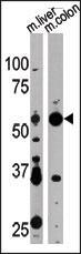
- Experimental details
- Western blot analysis of anti-APG4D Pab (Cat. #AP1811a) in mouse liver and colon tissue lysate. APG4D (arrow) was detected using the purified Pab.
- Primary Ab dilution
- 1:1000
- Submitted by
- Abcepta (provider)
- Main image
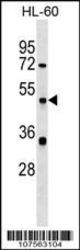
- Experimental details
- APG4D Antibody (N-term)(Cat. #AP1811a) western blot analysis in HL-60 cell line lysates (35ug/lane).This demonstrates the APG4D antibody detected the APG4D protein (arrow).
- Primary Ab dilution
- 1:1000

Supportive validation
- Submitted by
- Abcepta (provider)
- Main image
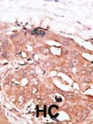
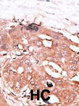
- Experimental details
- "Formalin-fixed and paraffin-embedded human cancer tissue reacted with the primary antibody, which was peroxidase-conjugated to the secondary antibody, followed by DAB staining. This data demonstrates the use of this antibody for immunohistochemistry; clinical relevance has not been evaluated. BC = breast carcinoma; HC = hepatocarcinoma."
- Primary Ab dilution
- 1:50~100