Explore
 Validate
 Learn
Learn Western blot
 Immunohistochemistry
Antibody data
- Antibody Data
- Antigen structure
- References [1]
- Comments [0]
- Validations
- Immunohistochemistry [1]
- Flow cytometry [2]
- Other assay [1]
Submit
Validation data
Reference
Comment
Report error
- Product number
- PA5-47375 - Provider product page

- Provider
- Invitrogen Antibodies
- Product name
- SPARCL1 Polyclonal Antibody
- Antibody type
- Polyclonal
- Antigen
- Recombinant full-length protein
- Description
- In direct ELISAs, approximately 15% cross-reactivity with recombinant human (rh) SPARC-like 1/SPARCL1 is observed and less than 1% cross-reactivity with rhSPARC is observed. Reconstitute at 0.2 mg/mL in sterile PBS.
- Reactivity
- Human
- Host
- Goat
- Isotype
- IgG
- Vial size
- 100 μg
- Concentration
- 0.2 mg/mL
- Storage
- -20°C, Avoid Freeze/Thaw Cycles
Submitted references Novel Combination of Surface Markers for the Reliable and Comprehensive Identification of Human Thymic Epithelial Cells by Flow Cytometry: Quantitation and Transcriptional Characterization of Thymic Stroma in a Pediatric Cohort.
Haunerdinger V, Moccia MD, Opitz L, Vavassori S, Dave H, Hauri-Hohl MM
Frontiers in immunology 2021;12:740047
Frontiers in immunology 2021;12:740047
No comments: Submit comment
Supportive validation
- Submitted by
- Invitrogen Antibodies (provider)
- Main image

- Experimental details
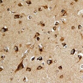
- Immunohistochemical analysis of SPARCL1 in immersion fixed paraffin-embedded sections of human brain. Samples were incubated in SPARCL1 polyclonal antibody (Product # PA5-47375) using a dilution of 15 µg/mL overnight at 4 °C. Before incubation with the primary antibody, tissue was subjected to heat-induced epitope retrieval using Antigen Retrieval Reagent-Basic . Tissue was stained using the Anti-Goat HRP-DAB Cell & Tissue Staining Kit (brown) and counterstained with hematoxylin (blue). Specific staining was localized to neurons.
Supportive validation
- Submitted by
- Invitrogen Antibodies (provider)
- Main image
- Experimental details
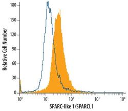
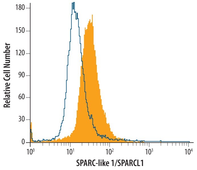
- Flow cytometry of SPARCL1 in HL‚60 human acute promyelocytic leukemia cell line. Samples were incubated in SPARCL1 polyclonal antibody (Product # PA5-47375) or control antibody followed by Phycoerythrin-conjugated Anti-Goat IgG Secondary Antibody. To facilitate intracellular staining, cells were fixed with paraformaldehyde and permeabilized with saponin.

- Submitted by
- Invitrogen Antibodies (provider)
- Main image
- Experimental details
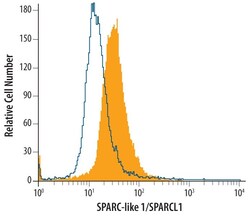
- Flow cytometry of SPARCL1 in HL‚60 human acute promyelocytic leukemia cell line. Samples were incubated in SPARCL1 polyclonal antibody (Product # PA5-47375) or control antibody followed by Phycoerythrin-conjugated Anti-Goat IgG Secondary Antibody. To facilitate intracellular staining, cells were fixed with paraformaldehyde and permeabilized with saponin.
Supportive validation
- Submitted by
- Invitrogen Antibodies (provider)
- Main image
- Experimental details
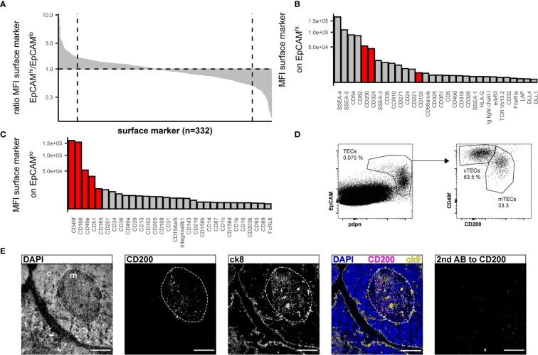
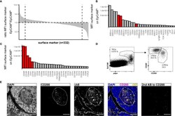
- Figure 2 Flow cytometry surface marker screen for markers distinguishing human cTECs and mTECs. (A) All screened surface markers plotted based on the ratio of MFI between CD45 - EpCAM high (putative mTECs) and CD45 - EpCAM low (putative cTECs). Dashed line: top 30 markers. (B, C) Top 30 surface markers from (A) with high EpCAM high /EpCAM low MFI ratio (B) or low EpCAM high /EpCAM low ratio (C) plotted by absolute MFI. Candidates that were chosen for further evaluation are marked in red. (D) Flow cytometry identification of TECs based on EpCAM and pdpn (left plot) and delineation of cTECs and mTECs based on CD49f and CD200 from live single cells from APC-enriched human thymus cell suspension. (E) Cryosections of human thymus. Nuclei are visualized with DAPI (blue). Cortex (c) and medulla (m) are distinguished based on density of nuclei (dashed line). Staining for CD200 (magenta). TECs are visualized with antibodies against ck8 (yellow). Exposure time for CD200 was adapted according to the staining with secondary antibody to CD200 (left panel). Scale bar 100 mum.