Explore
 Validate
 Learn
LearnAM32005PU-N
antibody from Acris Antibodies GmbH
Targeting: PTPRR
EC-PTP, PCPTP1, PTP-SL, PTPBR7, PTPRQ
 Western blot
 Immunoprecipitation
Antibody data
- Antibody Data
- Antigen structure
- References [0]
- Comments [0]
- Validations
- Western blot [2]
- Immunohistochemistry [1]
Submit
Validation data
Reference
Comment
Report error
- Product number
- AM32005PU-N - Provider product page

- Provider
- Acris Antibodies GmbH
- Proper citation
- Acris Antibodies GmbH Cat#AM32005PU-N, RRID:AB_11217985
- Product name
- anti PTPRR
- Antibody type
- Monoclonal
- Antigen
- Recombinant PTP-SL-GST (NCBI accession number BN000437, expression vector pGEX-2T), expressed in E.coli.
- Reactivity
- Mouse
- Host
- Mouse
- Isotype
- IgG
- Antibody clone number
- 6A6
- Vial size
- 0.1 mg
- Concentration
- 0.5 mg/ml (BSA concentration = 1%)
No comments: Submit comment
Supportive validation
- Submitted by
- Acris Antibodies GmbH (provider)
- Main image
- Experimental details
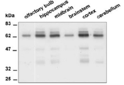
- Figure 2: Monoclonal antibody 6A6 immunoprecipitates from different brain regions, visualized on blot using STEP absorbed α-SL that is immunoreactive towards all PTPRR proteins.

- Submitted by
- Acris Antibodies GmbH (provider)
- Main image

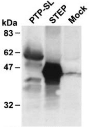
- Experimental details
- Figure 1: Neuro-2a cells were transiently transfected with PTP-SL or STEP expression plasmids, as indicated above the lanes. Mock transfected cells were included as negative controls. Lysates were directly subjected to Western blot analysis using monoclonal antibodies 6A6. In the PTP-SL Lane: at 60 kDa PTP-SL, at 42 and 37 kDa two PTPPBSγ isoforms.
Supportive validation
- Submitted by
- Acris Antibodies GmbH (provider)
- Main image

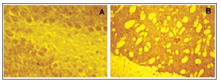
- Experimental details
- Figure 3: Immunolocalization of PTPRR protein in Mouse brain. Brain cryosections were stained using a mixture of three different monoclonalantibodies (1E3, 3E11 and 6A6) immunoreactive towards the common part inPTPRR isoforms and that cross-react with STEP. Positive staining is observed in the Purkinje cells of the cerebellum (A), both the neurones and neuropil of the striatum (B). The staining of the striatum reflects STEP immunoreactivity.