 Explore
 Validate
 Learn
Learn Immunohistochemistry
Antibody data
- Antibody Data
- Antigen structure
- References [1]
- Comments [0]
- Validations
- Immunohistochemistry [4]
Submit
Validation data
Reference
Comment
Report error
- Product number
- NBP2-13442 - Provider product page

- Provider
- Novus Biologicals
- Product name
- Rabbit Polyclonal TMEM106C Antibody
- Antibody type
- Polyclonal
- Description
- Immunogen affinity purified. Specificity of human TMEM106C antibody verified on a Protein Array containing target protein plus 383 other non-specific proteins.
- Reactivity
- Human
- Host
- Rabbit
- Isotype
- IgG
- Vial size
- 0.1 ml
- Storage
- Store at 4C short term. Aliquot and store at -20C long term. Avoid freeze-thaw cycles.
Submitted references Transmembrane protein 106C promotes the development of hepatocellular carcinoma.
Luo X, Han G, Lu R, Guan S, Wang Y, Ju L, Chen L, Shao J, Bian Z
Journal of cellular biochemistry 2020 Feb 9;
Journal of cellular biochemistry 2020 Feb 9;
No comments: Submit comment
Supportive validation
- Submitted by
- Novus Biologicals (provider)
- Main image
- Experimental details
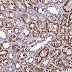
- Immunohistochemistry-Paraffin: TMEM106C Antibody [NBP2-13442] - Staining of human kidney shows strong granular cytoplasmic positivity in renal tubules.

- Submitted by
- Novus Biologicals (provider)
- Main image
- Experimental details
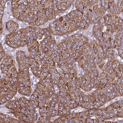
- Immunohistochemistry-Paraffin: TMEM106C Antibody [NBP2-13442] - Staining of human parathyroid gland shows high expression.

- Submitted by
- Novus Biologicals (provider)
- Main image

- Experimental details
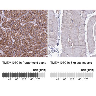
- Immunohistochemistry-Paraffin: TMEM106C Antibody [NBP2-13442] - Staining in human parathyroid gland and skeletal muscle tissues using anti-TMEM106C antibody. Corresponding TMEM106C RNA-seq data are presented for the same tissues.
- Submitted by
- Novus Biologicals (provider)
- Main image

- Experimental details
- Immunohistochemistry-Paraffin: TMEM106C Antibody [NBP2-13442] - Staining of human skeletal muscle shows low expression as expected.
