 Explore
 Validate
 Learn
Learn Western blot
 ELISA
ELISA Immunocytochemistry
Antibody data
- Antibody Data
- Antigen structure
- References [0]
- Comments [0]
- Validations
- Western blot [3]
- Immunocytochemistry [2]
- Immunoprecipitation [2]
Submit
Validation data
Reference
Comment
Report error
- Product number
- LS-C676847 - Provider product page

- Provider
- LSBio
- Product name
- HIST1H1C Antibody LS-C676847
- Antibody type
- Polyclonal
- Description
- Immunoaffinity purified
- Reactivity
- Human
- Host
- Rabbit
- Isotype
- IgG
- Storage
- Upon receipt, store at -20°C or -80°C. Avoid repeated freeze.
No comments: Submit comment
Enhanced validation
- Submitted by
- LSBio (provider)
- Enhanced method
- Genetic validation
- Main image

- Experimental details
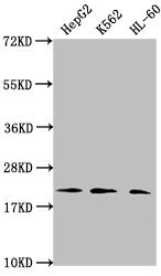
- Western Blot Positive WB detected in: HepG2 whole cell lysate, K562 whole cell lysate, HL-60 whole cell lysate All lanes: HIST1H1C antibody at 1:2000 Secondary Goat polyclonal to rabbit IgG at 1/40000 dilution Predicted band size: 22 kDa Observed band size: 22 kDa
- Submitted by
- LSBio (provider)
- Enhanced method
- Genetic validation
- Main image
- Experimental details
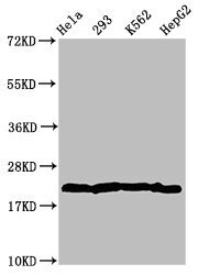
- Western Blot Positive WB detected in: Hela whole cell lysate, 293 whole cell lysate, K562 whole cell lysate, HepG2 whole cell lysate All Lanes: HIST1H1C antibody at 2.8µg/ml Secondary Goat polyclonal to rabbit IgG at 1/50000 dilution Predicted band size: 22 KDa Observed band size: 22 KDa
- Submitted by
- LSBio (provider)
- Main image

- Experimental details
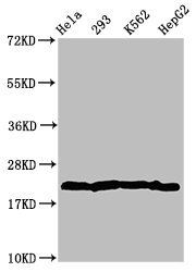
- Western Blot Positive WB detected in: Hela whole cell lysate, 293 whole cell lysate, K562 whole cell lysate, HepG2 whole cell lysate All Lanes: HIST1H1C antibody at 2.8µg/ml Secondary Goat polyclonal to rabbit IgG at 1/50000 dilution Predicted band size: 22 KDa Observed band size: 22 KDa

Supportive validation
- Submitted by
- LSBio (provider)
- Enhanced method
- Genetic validation
- Main image
- Experimental details
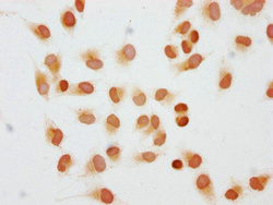
- Immunocytochemistry analysis of Di-methyl-HIST1H1C (K45) Antibody diluted at 1:10 and staining in Hela cells performed on a Leica BondTM system. The cells were fixed in 4% formaldehyde, permeabilized using 0.2% Triton X-100 and blocked with 10% normal goat serum 30min at RT. Then primary antibody (1% BSA) was incubated at 4°C overnight. The primary is detected by a biotinylated secondary antibody and visualized using an HRP conjugated SP system.

- Submitted by
- LSBio (provider)
- Main image

- Experimental details
- Immunocytochemistry analysis of Di-methyl-HIST1H1C (K45) Antibody diluted at 1:10 and staining in Hela cells performed on a Leica BondTM system. The cells were fixed in 4% formaldehyde, permeabilized using 0.2% Triton X-100 and blocked with 10% normal goat serum 30min at RT. Then primary antibody (1% BSA) was incubated at 4°C overnight. The primary is detected by a biotinylated secondary antibody and visualized using an HRP conjugated SP system.

Supportive validation
- Submitted by
- LSBio (provider)
- Enhanced method
- Genetic validation
- Main image
- Experimental details
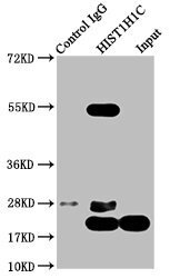
- Immunoprecipitating HIST1H1C in Hela whole cell lysate Lane 1: Rabbit control IgG instead of Di-methyl-HIST1H1C (K45) Antibody in Hela whole cell lysate.For western blotting, a HRP-conjugated Protein G antibody was used as the secondary antibody (1/2000) Lane 2: Di-methyl-HIST1H1C (K45) Antibody (5µg) + Hela whole cell lysate (1mg) Lane 3: Hela whole cell lysate (20µg)

- Submitted by
- LSBio (provider)
- Main image

- Experimental details
- Immunoprecipitating HIST1H1C in Hela whole cell lysate Lane 1: Rabbit control IgG instead of Di-methyl-HIST1H1C (K45) Antibody in Hela whole cell lysate.For western blotting, a HRP-conjugated Protein G antibody was used as the secondary antibody (1/2000) Lane 2: Di-methyl-HIST1H1C (K45) Antibody (5µg) + Hela whole cell lysate (1mg) Lane 3: Hela whole cell lysate (20µg)
