Explore
 Validate
 Learn
Learn Western blot
 Immunocytochemistry
Antibody data
- Antibody Data
- Antigen structure
- References [1]
- Comments [0]
- Validations
- Immunocytochemistry [4]
- Immunoprecipitation [1]
- Immunohistochemistry [3]
- Other assay [5]
Submit
Validation data
Reference
Comment
Report error
- Product number
- PA5-28622 - Provider product page

- Provider
- Invitrogen Antibodies
- Product name
- CrkL Polyclonal Antibody
- Antibody type
- Polyclonal
- Antigen
- Recombinant full-length protein
- Description
- Recommended positive controls: 293T, A431, HeLa, HepG2, mouse brain, U2OS. Predicted reactivity: Mouse (96%), Rat (96%), Zebrafish (81%), Xenopus laevis (87%), Rhesus Monkey (100%). Store product as a concentrated solution. Centrifuge briefly prior to opening the vial.
- Reactivity
- Human, Mouse, Rat
- Host
- Rabbit
- Isotype
- IgG
- Vial size
- 100 μL
- Concentration
- 0.33 mg/mL
- Storage
- Store at 4°C short term. For long term storage, store at -20°C, avoiding freeze/thaw cycles.
Submitted references The Effector TepP Mediates Recruitment and Activation of Phosphoinositide 3-Kinase on Early Chlamydia trachomatis Vacuoles.
Carpenter V, Chen YS, Dolat L, Valdivia RH
mSphere 2017 Jul-Aug;2(4)
mSphere 2017 Jul-Aug;2(4)
No comments: Submit comment
Supportive validation
- Submitted by
- Invitrogen Antibodies (provider)
- Main image

- Experimental details
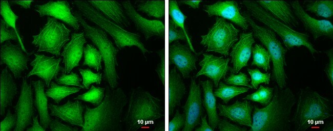
- Immunocytochemistry-Immunofluorescence analysis of CrkL was performed in HeLa cells fixed in 4% paraformaldehyde at RT for 15 min. Green: CrkL Polyclonal Antibody (Product # PA5-28622) diluted at 1:500. Blue: Hoechst 33342 staining. Scale bar = 10 µm.
- Submitted by
- Invitrogen Antibodies (provider)
- Main image
- Experimental details
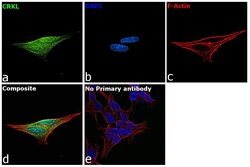
- Immunofluorescence analysis of Crk-like protein was performed using 70% confluent log phase SH-SY5Y cells. The cells were fixed with 4% paraformaldehyde for 10 minutes, permeabilized with 0.1% Triton™ X-100 for 15 minutes, and blocked with 2% BSA for 45 minutes at room temperature. The cells were labeled with CrkL Polyclonal Antibody (Product # PA5-28622) at 5 µg/mL in 0.1% BSA, incubated at 4 degree celsius overnight and then labeled with Goat anti-Rabbit IgG (H+L) Superclonal™ Recombinant Secondary Antibody, Alexa Fluor® 488 conjugate (Product # A27034), (1:2000 dilution), for 45 minutes at room temperature (Panel a: Green). Nuclei (Panel b:Blue) were stained with ProLong™ Diamond Antifade Mountant with DAPI (Product # P36962). F-actin (Panel c: Red) was stained with Rhodamine Phalloidin (Product # R415, 1:300 dilution). Panel d represents the merged image showing Cytoplasm localization. Panel e represents control cells with no primary antibody to assess background. The images were captured at 60x magnification.

- Submitted by
- Invitrogen Antibodies (provider)
- Main image

- Experimental details
- Immunocytochemistry-Immunofluorescence analysis of CrkL was performed in HeLa cells fixed in 4% paraformaldehyde at RT for 15 min. Green: CrkL Polyclonal Antibody (Product # PA5-28622) diluted at 1:500. Blue: Hoechst 33342 staining. Scale bar = 10 µm.

- Submitted by
- Invitrogen Antibodies (provider)
- Main image

- Experimental details
- Immunofluorescence analysis of Crk-like protein was performed using 70% confluent log phase SH-SY5Y cells. The cells were fixed with 4% paraformaldehyde for 10 minutes, permeabilized with 0.1% Triton™ X-100 for 15 minutes, and blocked with 2% BSA for 45 minutes at room temperature. The cells were labeled with CrkL Polyclonal Antibody (Product # PA5-28622) at 5 µg/mL in 0.1% BSA, incubated at 4 degree celsius overnight and then labeled with Goat anti-Rabbit IgG (Heavy Chain) Superclonal™ Recombinant Secondary Antibody, Alexa Fluor® 488 conjugate (Product # A27034), (1:2000 dilution), for 45 minutes at room temperature (Panel a: Green). Nuclei (Panel b:Blue) were stained with ProLong™ Diamond Antifade Mountant with DAPI (Product # P36962). F-actin (Panel c: Red) was stained with Rhodamine Phalloidin (Product # R415, 1:300 dilution). Panel d represents the merged image showing Cytoplasm localization. Panel e represents control cells with no primary antibody to assess background. The images were captured at 60x magnification.

Supportive validation
- Submitted by
- Invitrogen Antibodies (provider)
- Main image
- Experimental details
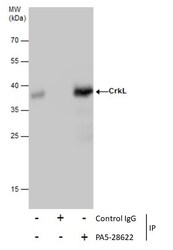
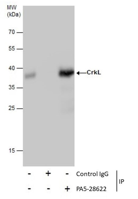
- Immunoprecipitation of CrkL was performed in 293T whole cell extracts using 5 µg of CrkL Polyclonal Antibody (Product # PA5-28622). Samples were transferred to a membrane and probed with CrkL Polyclonal Antibody as a primary antibody and an HRP-conjugated anti-Rabbit IgG was used as a secondary antibody.

Supportive validation
- Submitted by
- Invitrogen Antibodies (provider)
- Main image
- Experimental details
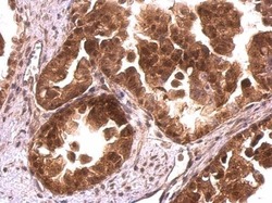
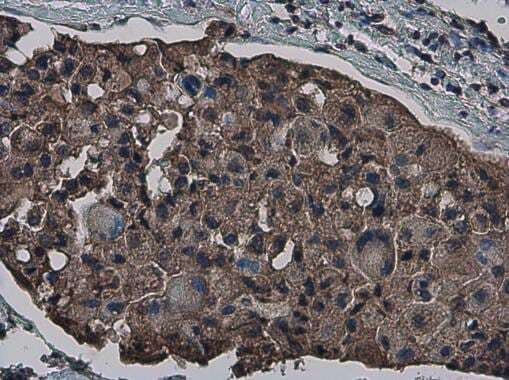
- CrkL Polyclonal Antibody detects CrkL protein at cytosol and nucleus on human ovarian carcinoma by immunohistochemical analysis. Sample: Paraffin-embedded human ovarian carcinoma. CrkL Polyclonal Antibody (Product # PA5-28622) dilution: 1:500. Antigen Retrieval: EDTA based buffer, pH 8.0, 15 min.

- Submitted by
- Invitrogen Antibodies (provider)
- Main image
- Experimental details
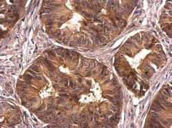
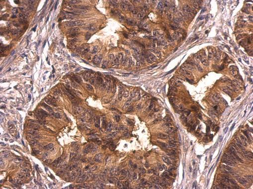
- CrkL Polyclonal Antibody detects CrkL protein at cytosol on human colon carcinoma by immunohistochemical analysis. Sample: Paraffin-embedded human colon carcinoma. CrkL Polyclonal Antibody (Product # PA5-28622) dilution: 1:500. Antigen Retrieval: EDTA based buffer, pH 8.0, 15 min.
- Submitted by
- Invitrogen Antibodies (provider)
- Main image
- Experimental details
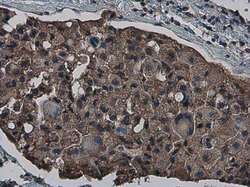
- Immunohistochemistry (Paraffin) analysis of CrkL was performed in paraffin-embedded human breast carcinoma tissue using CrkL Polyclonal Antibody (Product # PA5-28622) at a dilution of 1:500.
Supportive validation
- Submitted by
- Invitrogen Antibodies (provider)
- Main image
- Experimental details
- NULL

- Submitted by
- Invitrogen Antibodies (provider)
- Main image

- Experimental details
- Immunoprecipitation of CrkL was performed in 293T whole cell extracts using 5 µg of CrkL Polyclonal Antibody (Product # PA5-28622). Samples were transferred to a membrane and probed with CrkL Polyclonal Antibody as a primary antibody and an HRP-conjugated anti-Rabbit IgG was used as a secondary antibody.
- Submitted by
- Invitrogen Antibodies (provider)
- Main image

- Experimental details
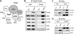
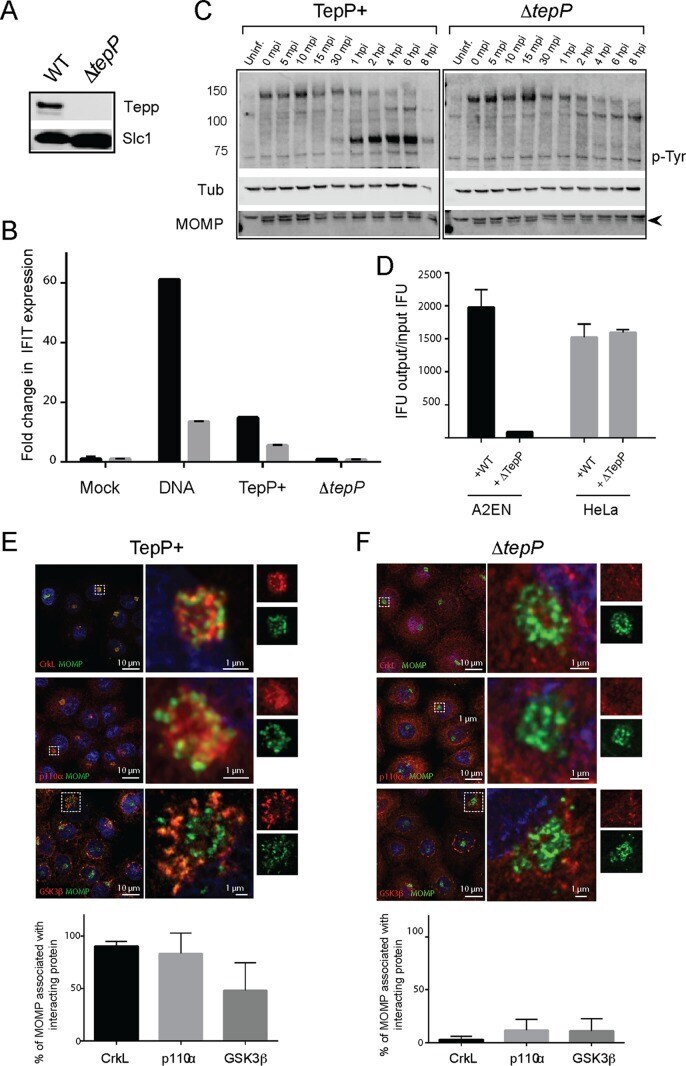
- FIG 2 TepP is required for C. trachomatis replication in A2EN cells and to recruit CrkL and PI3K to nascent inclusions. (A to D) Phenotypic characterization of a C. trachomatis Delta tepP :: bla insertional mutant. (A) Immunoblot analysis of TepP expression in HeLa cells infected for 24 h with the indicated strains. Slc1, loading control. (B) A2EN cells were infected with CTL2 or Delta tepP :: bla bacteria, and the levels of IFIT1 (black) and IFIT2 (gray) transcripts from the strains described in the panel A legend were assessed by quantitative RT-PCR. DNA transfection was used as a positive control for the induction of IFIT genes. (C) Time course of appearance of major tyrosine-phosphorylated (p-Tyr) proteins in A2EN cell infected with CTL2 or Delta tepP :: bla insertional mutants or left uninfected (Uninf.). MOMP, bacterial major outer membrane protein (arrowhead); Tub, tubulin. (D) The replication potential of Delta tepP :: bla mutants was assessed in HeLa and A2EN cells by the generation of inclusion-forming units (IFU). (E and F) Subcellular localization of CrkL, PI3K (p110alpha), and GSK-3beta in Chlamydia -infected cells. A2EN cells were infected with CTL2 or Delta tepP :: bla bacteria for 4 h and immunostained by anti-MOMP (green), anti-CrkL, anti-p110alpha, or anti-GSK-3beta (red). Hoechst (blue) was used to detect DNA. Quantification of Chlamydia (MOMP) and p110alpha, CrkL, and GSK-3beta colocalization was performed on a single-cell basis ( n = 20 and 30 cells/repli
- Submitted by
- Invitrogen Antibodies (provider)
- Main image
- Experimental details
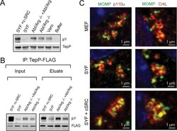
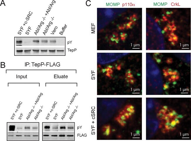
- FIG 3 TepP is phosphorylated by Src kinases. (A) In vitro phosphorylation reactions were performed by incubating recombinant 6xHis-TepP with ATP and cell lysates derived from the indicated cell lines. The degree of TepP phosphorylation was assessed by immunoblotting with anti-pY antibodies after reisolation of TepP on nickel beads. TepP was phosphorylated under all conditions except after incubation with SYF cell lysates and the buffer-only control. (B) TepP-FLAG immunoprecipitations from cell lines infected with CTLM062G1 expressing TepP-FLAG. TepP phosphorylation was assessed after IP with anti-FLAG antibodies and immunoblotting with anti-pY antibodies. Note the decreased levels of phosphorylation in Src, Yes, and Fyn-deficient cells. (C) Mouse embryo fibroblast (MEF), SYF, and SYF-plus-c-Src cell lines were infected for 4 h with C. trachomatis at an MOI of 20, fixed, and stained. Infected cells were immunostained by anti-MOMP (green), anti-CrkL, or anti-p110alpha (red) antibodies.
- Submitted by
- Invitrogen Antibodies (provider)
- Main image
- Experimental details
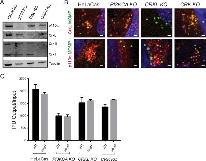
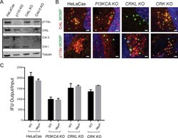
- FIG 4 PI3K and CrkL independently associate with early inclusions. (A) HeLa cells stably expressing Cas9 (HeLa-Cas) were transduced with sgRNAs specific to PIK3KCA , CRKL , and CRKI-II to generate gene-edited cell lines lacking expression of the respective target proteins. Loss of protein expression for each cell line was verified by immunoblot analysis using anti-p110alpha, anti-CrkL, and anti-Crk I/II antibodies. Tubulin levels were used as positive controls. There was a low level of cross-reactivity between CrkL and CrkII antibodies. (B) CrkL and p110alpha are recruited to early inclusions formed in PI3KCA and CRKL gene-edited knockout cells (KO), respectively. HeLa-Cas cells and their edited derivatives were infected with CTL2 for 4 h, fixed, and immunostained with anti-MOMP and anti-p110alpha or anti-CrkL (red) antibodies. Host and bacterial DNA were detected with Hoechst (blue). (C) Replication of WT and TepP-deficient C. trachomatis in PI3KCA , CRKL , and CRK gene-edited knockout cells (KO).