 Explore
 Validate
 Learn
Learn Western blot
 Immunocytochemistry
Antibody data
- Antibody Data
- Antigen structure
- References [0]
- Comments [0]
- Validations
- Western blot [3]
- Immunohistochemistry [6]
Submit
Validation data
Reference
Comment
Report error
- Product number
- LS-C156268 - Provider product page

- Provider
- LSBio
- Product name
- ALDH6A1 Antibody LS-C156268
- Antibody type
- Monoclonal
- Description
- Protein G purified
- Reactivity
- Human
- Host
- Mouse
- Isotype
- IgG
- Storage
- Maintain refrigerated at 2°C to 8°C for up to 6 months. For long term storage store at -20°C.
No comments: Submit comment
Enhanced validation
- Submitted by
- LSBio (provider)
- Enhanced method
- Genetic validation
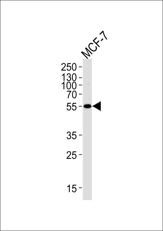
- Main image

- Experimental details
- ALDH6A1 Antibody western blot of MCF-7 cell lysate (35 ug/lane). This demonstrates that the ALDH6A1 antibody detected ALDH6A1 protein (arrow).
- Submitted by
- LSBio (provider)
- Enhanced method
- Genetic validation
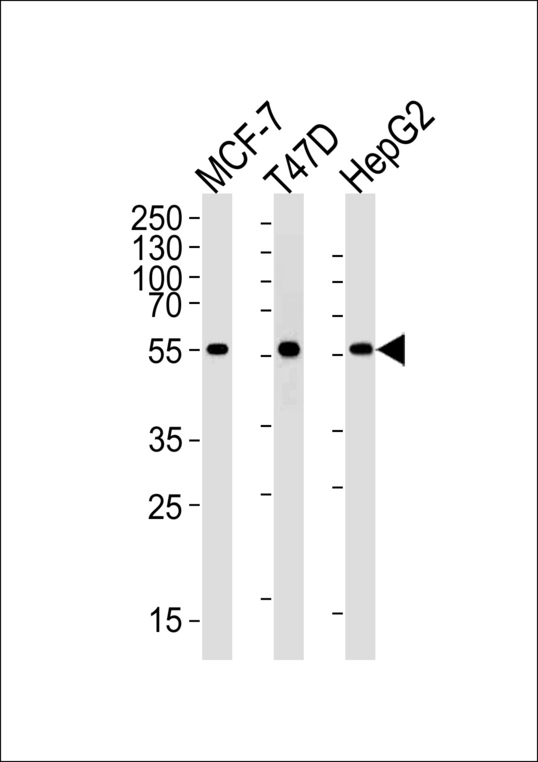
- Main image

- Experimental details
- ALDH6A1 Antibody western blot of MCF-7, T47D, HepG2 cell lysates (35 ug/lane). This demonstrates that the ALDH6A1 antibody detected ALDH6A1 protein (arrow).
- Submitted by
- LSBio (provider)
- Enhanced method
- Genetic validation
- Main image
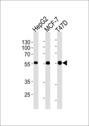
- Experimental details
- ALDH6A1 Antibody western blot of HepG2, MCF-7,T47D cell line lysates (35 ug/lane). The ALDH6A1 antibody detected the ALDH6A1 protein (arrow).

Supportive validation
- Submitted by
- LSBio (provider)
- Enhanced method
- Genetic validation
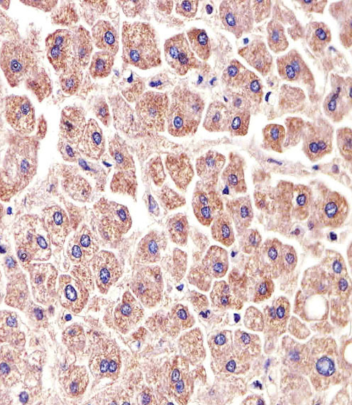
- Main image

- Experimental details
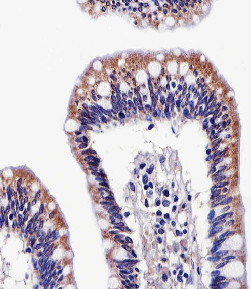
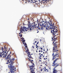
- Immunohistochemical of paraffin-embedded H.colon section using ALDH6A1 Antibody. Antibody was diluted at 1:25 dilution. A peroxidase-conjugated goat anti-rabbit IgG at 1:400 dilution was used as the secondary antibody, followed by DAB staining.

- Submitted by
- LSBio (provider)
- Enhanced method
- Genetic validation
- Main image

- Experimental details
- Immunohistochemical of paraffin-embedded H.liver section using ALDH6A1 Antibody. Antibody was diluted at 1:25 dilution. A peroxidase-conjugated goat anti-rabbit IgG at 1:400 dilution was used as the secondary antibody, followed by DAB staining.
- Submitted by
- LSBio (provider)
- Enhanced method
- Genetic validation
- Main image

- Experimental details
- Immunohistochemical of paraffin-embedded H.colon section using ALDH6A1 Antibody. Antibody was diluted at 1:25 dilution. A peroxidase-conjugated goat anti-rabbit IgG at 1:400 dilution was used as the secondary antibody, followed by DAB staining.
- Submitted by
- LSBio (provider)
- Enhanced method
- Genetic validation
- Main image

- Experimental details
- Immunohistochemical of paraffin-embedded H.liver section using ALDH6A1 Antibody. Antibody was diluted at 1:25 dilution. A peroxidase-conjugated goat anti-rabbit IgG at 1:400 dilution was used as the secondary antibody, followed by DAB staining.

- Submitted by
- LSBio (provider)
- Enhanced method
- Genetic validation
- Main image
- Experimental details
- Immunohistochemical of paraffin-embedded H.colon section using ALDH6A1 Antibody. Antibody was diluted at 1:25 dilution. A peroxidase-conjugated goat anti-rabbit IgG at 1:400 dilution was used as the secondary antibody, followed by DAB staining.

- Submitted by
- LSBio (provider)
- Enhanced method
- Genetic validation
- Main image

- Experimental details
- Immunohistochemical of paraffin-embedded H.liver section using ALDH6A1 Antibody. Antibody was diluted at 1:25 dilution. A peroxidase-conjugated goat anti-rabbit IgG at 1:400 dilution was used as the secondary antibody, followed by DAB staining.
