Explore
 Validate
 Learn
Learn Western blot
Antibody data
- Antibody Data
- Antigen structure
- References [1]
- Comments [0]
- Validations
- Western blot [1]
- Immunocytochemistry [1]
- Immunohistochemistry [2]
Submit
Validation data
Reference
Comment
Report error
- Product number
- MAB4751 - Provider product page

- Provider
- R&D Systems
- Product name
- Human/Mouse Wnt-4 Antibody
- Antibody type
- Monoclonal
- Description
- Protein A or G purified from hybridoma culture supernatant. This antibody detects human and mouse Wnt-4 in direct ELISAs and Western blots. In direct ELISAs, no cross-reactivity with recombinant mouse (rm) Wnt-1, 2b, 5a, 5b, 8a, 8b, 9b, 10a, 10b, 11, 16, recombinant human (rh) Wnt-2, 3a, 7a, or 7b is observed.
- Reactivity
- Human, Mouse
- Host
- Rat
- Conjugate
- Unconjugated
- Antigen sequence
NP_033549- Isotype
- IgG
- Antibody clone number
- 55025
- Vial size
- 100 ug
- Concentration
- LYOPH
- Storage
- Use a manual defrost freezer and avoid repeated freeze-thaw cycles. 12 months from date of receipt, -20 to -70 °C as supplied. 1 month, 2 to 8 °C under sterile conditions after reconstitution. 6 months, -20 to -70 °C under sterile conditions after reconstitution.
Submitted references Restoration of WNT4 inhibits cell growth in leukemia-derived cell lines.
García-Castro B, Alvarez-Zavala M, Riveros-Magaña AR, Ortíz-Lazareno PC, Ratkovich-González S, Hernández-Flores G, Bravo-Cuellar A, Jave-Suarez LF, Aguilar-Lemarroy A
BMC cancer 2013 Nov 25;13:557
BMC cancer 2013 Nov 25;13:557
No comments: Submit comment
Supportive validation
- Submitted by
- R&D Systems (provider)
- Main image
- Experimental details
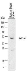
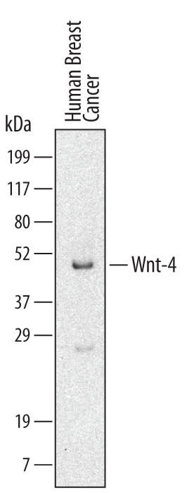
- Detection of Human Wnt-4 by Western Blot. Western blot shows lysates of human breast cancer tissue. PVDF membrane was probed with 2 µg/mL Rat Anti-Human/Mouse Wnt-4 Monoclonal Antibody (Catalog # MAB4751) followed by HRP-conjugated Anti-Rat IgG Secondary Antibody (Catalog # HAF005). A specific band for Wnt-4 was detected at approximately 39 kDa (as indicated). This experiment was conducted under reducing conditions and using Immunoblot Buffer Group 1.
Supportive validation
- Submitted by
- R&D Systems (provider)
- Main image
- Experimental details
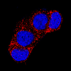
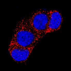
- Wnt-4 in MCF-7 Human Cell Line. Wnt-4 was detected in immersion fixed MCF-7 human breast cancer cell line using Rat Anti-Human/Mouse Wnt-4 Monoclonal Antibody (Catalog # MAB4751) at 3 µg/mL for 3 hours at room temperature. Cells were stained using the NorthernLights™ 557-conjugated Anti-Rat IgG Secondary Antibody (red; Catalog # NL013) and counterstained with DAPI (blue). Specific staining was localized to cytoplasm. View our protocol for Fluorescent ICC Staining of Cells on Coverslips.
Supportive validation
- Submitted by
- R&D Systems (provider)
- Main image
- Experimental details
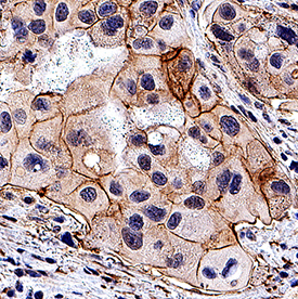
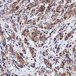
- Wnt-4 in Human Breast Cancer Tissue. Wnt-4 was detected in immersion fixed paraffin-embedded sections of human breast cancer tissue using Rat Anti-Human/Mouse Wnt-4 Monoclonal Antibody (Catalog # MAB4751) at 1.7 µg/mL overnight at 4 °C. Tissue was stained using the Anti-Rat HRP-DAB Cell & Tissue Staining Kit (brown; Catalog # CTS017) and counterstained with hematoxylin (blue). Specific staining was localized to cytoplasm in cancer cells. View our protocol for Chromogenic IHC Staining of Paraffin-embedded Tissue Sections.

- Submitted by
- R&D Systems (provider)
- Main image
- Experimental details
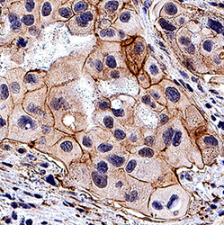
- Wnt-4 in Human Breast Cancer Tissue. Wnt-4 was detected in immersion fixed paraffin-embedded sections of human breast cancer tissue using Rat Anti-Human/Mouse Wnt-4 Monoclonal Antibody (Catalog # MAB4751) at 5 µg/mL for 1 hour at room temperature followed by incubation with the Anti-Rat IgG VisUCyte™ HRP Polymer Antibody (Catalog # VC005). Tissue was stained using DAB (brown) and counterstained with hematoxylin (blue). Specific staining was localized to plasma membrane. View our protocol for IHC Staining with VisUCyte HRP Polymer Detection Reagents.