Explore
 Validate
 Learn
LearnMA5-15796
antibody from Invitrogen Antibodies
Targeting: CARM1
PRMT4
 Western blot
 ELISA
ELISA Immunocytochemistry Immunohistochemistry Flow cytometry Chromatin Immunoprecipitation
Antibody data
- Antibody Data
- Antigen structure
- References [0]
- Comments [0]
- Validations
- Immunocytochemistry [4]
- Immunohistochemistry [1]
- Flow cytometry [2]
- Chromatin Immunoprecipitation [2]
Submit
Validation data
Reference
Comment
Report error
- Product number
- MA5-15796 - Provider product page

- Provider
- Invitrogen Antibodies
- Product name
- PRMT4 Monoclonal Antibody (3H2)
- Antibody type
- Monoclonal
- Antigen
- Purifed from natural sources
- Description
- MA5-15796 targets CARM1 in indirect ELISA, FACS, IF, IHC, and WB applications and shows reactivity with Human, Non-human primate, Mouse and Rat samples. The MA5-15796 immunogen is purified recombinant fragment of human CARM1 expressed in E. Coli. MA5-15796 detects CARM1 which has a predicted molecular weight of approximately 65kDa.
- Reactivity
- Human, Mouse, Rat
- Host
- Mouse
- Isotype
- IgG
- Antibody clone number
- 3H2
- Vial size
- 100 μL
- Concentration
- Conc. not determined
- Storage
- Store at 4°C short term. For long term storage, store at -20°C, avoiding freeze/thaw cycles.
No comments: Submit comment
Supportive validation
- Submitted by
- Invitrogen Antibodies (provider)
- Main image
- Experimental details
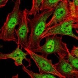
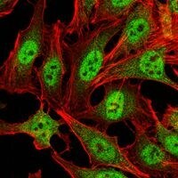
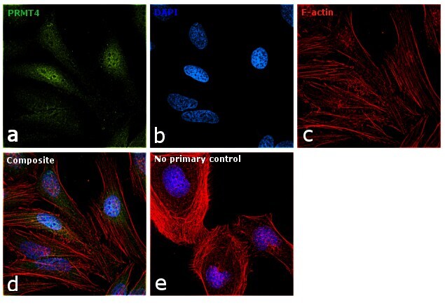
- Immunofluorescence analysis of HeLa cells using CARM1 monoclonal antibody (Product # MA5-15796) (Green). Red: actin filaments have been labeled with phalloidin.
- Submitted by
- Invitrogen Antibodies (provider)
- Main image
- Experimental details
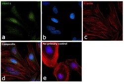
- Immunofluorescence analysis of PRMT4 was performed using 70% confluent log phase HeLa cells. The cells were fixed with 4% paraformaldehyde for 10 minutes, permeabilized with 0.1% Triton™ X-100 for 15 minutes, and blocked with 1% BSA for 1 hour at room temperature. The cells were labeled with PRMT4 Monoclonal Antibody (3H2) (Product # MA5-15796) at 1:200 dilution in 0.1% BSA, incubated at 4 degree Celsius overnight and then labeled with Goat anti-Mouse IgG (H+L) Superclonal™ Secondary Antibody, Alexa Fluor® 488 conjugate (Product #A28175) at a dilution of 1:2000 for 45 minutes at room temperature (Panel a: green). Nuclei (Panel b: blue) were stained with ProLong™ Diamond Antifade Mountant with DAPI (Product # P36962). F-actin (Panel c: red) was stained with Rhodamine Phalloidin (Product # R415). Panel d represents the merged image showing Nuclear localization. Panel e represents control cells with no primary antibody to assess background. The images were captured at 60X magnification.

- Submitted by
- Invitrogen Antibodies (provider)
- Main image

- Experimental details
- Immunofluorescence analysis of HeLa cells using CARM1 monoclonal antibody (Product # MA5-15796) (Green). Red: actin filaments have been labeled with phalloidin.

- Submitted by
- Invitrogen Antibodies (provider)
- Main image

- Experimental details
- Immunofluorescence analysis of PRMT4 was performed using 70% confluent log phase HeLa cells. The cells were fixed with 4% paraformaldehyde for 10 minutes, permeabilized with 0.1% Triton™ X-100 for 15 minutes, and blocked with 1% BSA for 1 hour at room temperature. The cells were labeled with PRMT4 Monoclonal Antibody (3H2) (Product # MA5-15796) at 1:200 dilution in 0.1% BSA, incubated at 4 degree Celsius overnight and then labeled with Goat anti-Mouse IgG (H+L) Superclonal™ Secondary Antibody, Alexa Fluor® 488 conjugate (Product #A28175) at a dilution of 1:2000 for 45 minutes at room temperature (Panel a: green). Nuclei (Panel b: blue) were stained with ProLong™ Diamond Antifade Mountant with DAPI (Product # P36962). F-actin (Panel c: red) was stained with Rhodamine Phalloidin (Product # R415). Panel d represents the merged image showing Nuclear localization. Panel e represents control cells with no primary antibody to assess background. The images were captured at 60X magnification.
Supportive validation
- Submitted by
- Invitrogen Antibodies (provider)
- Main image
- Experimental details
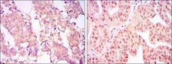
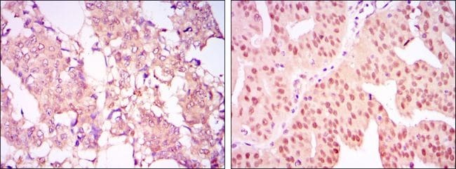
- Immunohistochemical analysis of paraffin-embedded breast cancer tissues (left) and ovarian cancer tissues (right) using CARM1 monoclonal antibody (Product # MA5-15796) followed with DAB staining.
Supportive validation
- Submitted by
- Invitrogen Antibodies (provider)
- Main image

- Experimental details
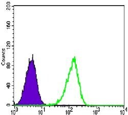
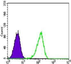
- Flow cytometric analysis of Lovo cells using CARM1 monoclonal antibody (Product # MA5-15796) (green) and negative control (purple).
- Submitted by
- Invitrogen Antibodies (provider)
- Main image
- Experimental details
- Flow cytometric analysis of Lovo cells using CARM1 monoclonal antibody (Product # MA5-15796) (green) and negative control (purple).

Supportive validation
- Submitted by
- Invitrogen Antibodies (provider)
- Main image

- Experimental details
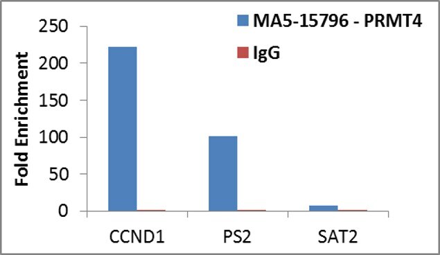
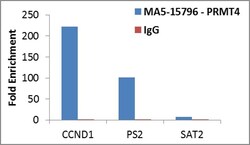
- Enrichment of endogenous PRMT4 protein at specific gene loci using Anti-PRMT4 Antibody: Chromatin Immunoprecipitation (ChIP) was performed using Anti-PRMT4 Monoclonal Antibody (Product # MA5-15796, 8 ul) on sheared chromatin from 2 million HCT 116 cells using the MAGnify ChIP system kit (Product # 49-2024). Normal Rabbit IgG was used as a negative IP control. The purified DNA was analyzed by qPCR with PCR primer pairs over the promoters of CCND1 and PS2 (positive) and SAT2 satellite repeats (negative). Data is presented as fold enrichment of the antibody signal versus the negative control IgG using the comparative CT method.

- Submitted by
- Invitrogen Antibodies (provider)
- Main image
- Experimental details
- Enrichment of endogenous PRMT4 protein at specific gene loci using Anti-PRMT4 Antibody: Chromatin Immunoprecipitation (ChIP) was performed using Anti-PRMT4 Monoclonal Antibody (Product # MA5-15796, 8 ul) on sheared chromatin from 2 million HCT 116 cells using the MAGnify ChIP system kit (Product # 49-2024). Normal Rabbit IgG was used as a negative IP control. The purified DNA was analyzed by qPCR with PCR primer pairs over the promoters of CCND1 and PS2 (positive) and SAT2 satellite repeats (negative). Data is presented as fold enrichment of the antibody signal versus the negative control IgG using the comparative CT method.