 Explore
 Validate
 Learn
Learn Western blot
 Other assay
Antibody data
- Antibody Data
- Antigen structure
- References [1]
- Comments [0]
- Validations
- Other assay [2]
Submit
Validation data
Reference
Comment
Report error
- Product number
- PA5-69987 - Provider product page

- Provider
- Invitrogen Antibodies
- Product name
- GID4 Polyclonal Antibody
- Antibody type
- Polyclonal
- Antigen
- Synthetic peptide
- Description
- This target displays homology in the following species: Cow: 100%; Dog: 100%; Horse: 100%; Human: 100%; Mouse: 100%; Rabbit: 100%; Rat: 100%; Zebrafish: 100%
- Reactivity
- Human
- Host
- Rabbit
- Isotype
- IgG
- Vial size
- 100 μL
- Concentration
- 0.5 mg/mL
- Storage
- -20°C, Avoid Freeze/Thaw Cycles
Submitted references The human GID complex engages two independent modules for substrate recruitment.
Mohamed WI, Park SL, Rabl J, Leitner A, Boehringer D, Peter M
EMBO reports 2021 Nov 4;22(11):e52981
EMBO reports 2021 Nov 4;22(11):e52981
No comments: Submit comment
Supportive validation
- Submitted by
- Invitrogen Antibodies (provider)
- Main image

- Experimental details
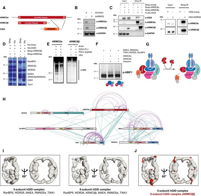
- Figure 4 ARMC8alpha but not ARMC8beta recruits GID4 to the core complex in an assembly that does not prevent binding of the WDR26/RanBP9 module Schematic representation of ARMC8alpha (Q8IUR7-1), ARMC8beta (Q8IUR7-6), and GID4 (Q8IVV7-1) proteins. The binding site of GID4 and the C-terminus of ARMC8alpha is indicated. Western blot analysis showing the levels of ARMC8alpha and ARMC8beta in HeLa Kyoto cells treated for 72 h with control siRNA or siRNA pools against ARMC8 ( n = 3). Transiently expressed FLAG-GID4 and HSS-tagged ARMC8 isoforms (alpha or beta) in HEK-293T cells. The presence of GID4 in isoform-specific ARMC8 immunoprecipitates was visualized by immunoblotting (left panels). The right panel shows a Western blot of transiently expressed and immunoprecipitated HSS-WDR26 from HEK-293T cells, and the presence of endogenous ARMC8 isoforms (alpha or beta) was probed by immunoblotting ( n = 2). Baculoviral co-expression in Sf9 cells of the 5-subunit hGID complex (5mer; His-RanBP9, His-WDR26, FLAG-MAEA, His-RMND5a, and His-TWA1) along with His-GID4 in the presence of Strep-ARMC8alpha or Strep-ARMC8beta. Strep- or His-pulldowns revealed the presence of GID4 in ARMC8alpha, but not in ARMC8beta, complexes ( n = 3). Immunoblot analysis of in vitro ubiquitinated GID4-hGID complexes containing either ARMC8alpha or ARMC8beta (6mer; ARMC8, RanBP9, WDR26, MAEA, RMND5a, and TWA1). Where indicated, the reaction was carried out in the presence of 20-fold molar excess of the PGLV GID4-b
- Submitted by
- Invitrogen Antibodies (provider)
- Main image
- Experimental details
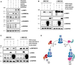
- Figure 1 The hGID complex uses distinct substrate modules to target different substrates A Immunoblot of cell extracts following depletion of WDR26, RanBP9, ARMC8, and GID4 using pools of siRNAs for 72 h in HeLa Kyoto cells. Endogenous levels of the indicated proteins were monitored by Western blotting ( n = 3). B, C Western blotting of samples after ectopic overexpression of HBP1 (B) or ZMYND19 (C) alone, or together with WDR26 or GID4 in HEK-293T cells. HBP1 and ZMYND19 levels were monitored after treatment of MG132 or DMSO for 10-12 h ( n = 3). D Schematic representation visualizing the hGID E3 ligase complex using two distinct modules for substrate recruitment. Source data are available online for this figure.
