Explore
 Validate
 Learn
Learn Western blot
Antibody data
- Antibody Data
- Antigen structure
- References [1]
- Comments [0]
- Validations
- Western blot [1]
- Immunocytochemistry [2]
- Immunohistochemistry [11]
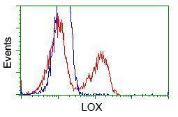
- Flow cytometry [1]
Submit
Validation data
Reference
Comment
Report error
- Product number
- GTX84182 - Provider product page

- Provider
- GeneTex
- Proper citation
- GeneTex Cat#GTX84182, RRID:AB_10734013
- Product name
- LOX antibody [6B11]
- Antibody type
- Monoclonal
- Reactivity
- Human
- Host
- Mouse
Submitted references Hypoxia Inducible Factors Modify Collagen I Fibers in MDA-MB-231 Triple Negative Breast Cancer Xenografts.
Goggins E, Kakkad S, Mironchik Y, Jacob D, Wildes F, Krishnamachary B, Bhujwalla ZM
Neoplasia (New York, N.Y.) 2018 Feb;20(2):131-139
Neoplasia (New York, N.Y.) 2018 Feb;20(2):131-139
No comments: Submit comment
Supportive validation
- Submitted by
- GeneTex (provider)
- Main image
- Experimental details
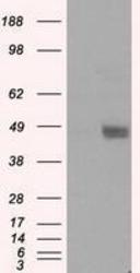
- HEK293T cells were transfected with the pCMV6-ENTRY control (Left lane) or pCMV6-ENTRY LOX (Right lane) cDNA for 48 hrs and lysed. Equivalent amounts of cell lysates (5 ug per lane) were separated by SDS-PAGE and immunoblotted with anti-LOX.
Supportive validation
- Submitted by
- GeneTex (provider)
- Main image
- Experimental details
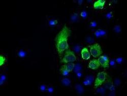
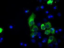
- Anti-LOX mouse monoclonal antibody (GTX84182) immunofluorescent staining of COS7 cells transiently transfected with LOX
- Submitted by
- GeneTex (provider)
- Main image

- Experimental details
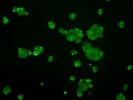
- Immunofluorescent staining of HepG2 cells using anti-LOX mouse monoclonal antibody (GTX84182).
Supportive validation
- Submitted by
- GeneTex (provider)
- Main image

- Experimental details
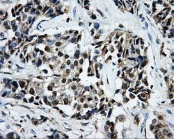
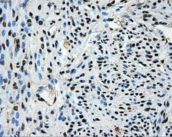
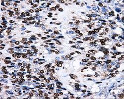
- Immunohistochemical staining of paraffin-embedded Adenocarcinoma of breast tissue using anti-LOX mouse monoclonal antibody. (GTX84182, Dilution 1:50)
- Submitted by
- GeneTex (provider)
- Main image

- Experimental details
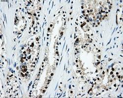
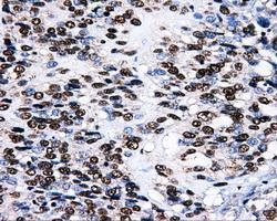
- Immunohistochemical staining of paraffin-embedded prostate tissue using anti-LOXmouse monoclonal antibody. (GTX84182, Dilution 1:50)

- Submitted by
- GeneTex (provider)
- Main image
- Experimental details
- Immunohistochemical staining of paraffin-embedded Carcinoma of prostate tissue using anti-LOXmouse monoclonal antibody. (GTX84182, Dilution 1:50)

- Submitted by
- GeneTex (provider)
- Main image
- Experimental details
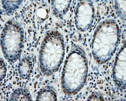
- Immunohistochemical staining of paraffin-embedded colon tissue using anti-LOXmouse monoclonal antibody. (GTX84182, Dilution 1:50)

- Submitted by
- GeneTex (provider)
- Main image

- Experimental details
- Immunohistochemical staining of paraffin-embedded endometrium tissue using anti-LOXmouse monoclonal antibody. (GTX84182, Dilution 1:50)
- Submitted by
- GeneTex (provider)
- Main image
- Experimental details
- Immunohistochemical staining of paraffin-embedded thyroid tissue using anti-LOXmouse monoclonal antibody. (GTX84182, Dilution 1:50)

- Submitted by
- GeneTex (provider)
- Main image
- Experimental details
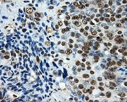
- Immunohistochemical staining of paraffin-embedded Adenocarcinoma of ovary tissue using anti-LOXmouse monoclonal antibody. (GTX84182, Dilution 1:50)

- Submitted by
- GeneTex (provider)
- Main image
- Experimental details
- Immunohistochemical staining of paraffin-embedded Carcinoma of bladder tissue using anti-LOXmouse monoclonal antibody. (GTX84182, Dilution 1:50)
- Submitted by
- GeneTex (provider)
- Main image

- Experimental details
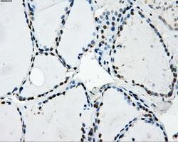
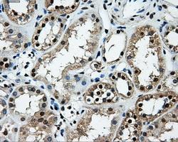
- Immunohistochemical staining of paraffin-embedded Kidney tissue using anti-LOXmouse monoclonal antibody. (GTX84182, Dilution 1:50)
- Submitted by
- GeneTex (provider)
- Main image

- Experimental details
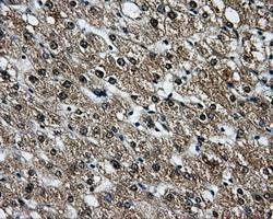
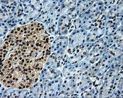
- Immunohistochemical staining of paraffin-embedded liver tissue using anti-LOXmouse monoclonal antibody. (GTX84182, Dilution 1:50)
- Submitted by
- GeneTex (provider)
- Main image

- Experimental details
- Immunohistochemical staining of paraffin-embedded pancreas tissue using anti-LOXmouse monoclonal antibody. (GTX84182, Dilution 1:50)
Supportive validation
- Submitted by
- GeneTex (provider)
- Main image
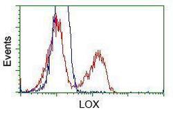
- Experimental details
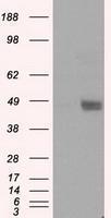
- HEK293T cells transfected with either pCMV6-ENTRY LOX