Explore
 Validate
 Learn
Learn Western blot
 Immunocytochemistry
Antibody data
- Antibody Data
- Antigen structure
- References [4]
- Comments [0]
- Validations
- Western blot [3]
- Flow cytometry [1]
Submit
Validation data
Reference
Comment
Report error
- Product number
- NBP1-30327 - Provider product page

- Provider
- Novus Biologicals
- Proper citation
- Novus Cat#NBP1-30327, RRID:AB_2037084
- Product name
- Rabbit Polyclonal LOX propeptide Antibody
- Antibody type
- Polyclonal
- Description
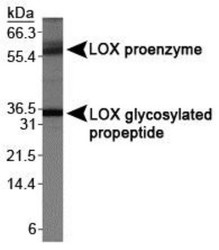
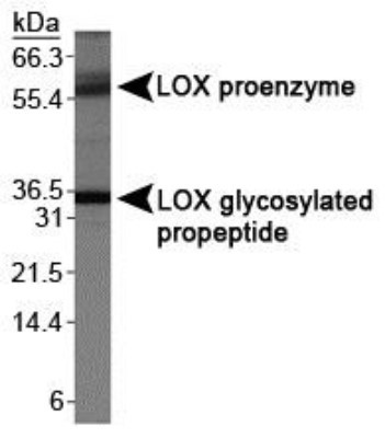
- Immunogen affinity purified. This antibody recognizes the glycosylated propeptide form (~35 kDa) and the proenzyme form (~50 kDa).
- Reactivity
- Human, Rat
- Host
- Rabbit
- Isotype
- IgG
- Vial size
- 0.1 ml
- Concentration
- 1 mg/ml
- Storage
- Store at 4C short term. Aliquot and store at -20C long term. Avoid freeze-thaw cycles.
Submitted references Prostaglandin E2 inhibits elastogenesis in the ductus arteriosus via EP4 signaling.
Inhibition of CIN85-mediated invasion by a novel SH3 domain binding motif in the lysyl oxidase propeptide.
The Ras signaling inhibitor LOX-PP interacts with Hsp70 and c-Raf to reduce Erk activation and transformed phenotype of breast cancer cells.
The lysyl oxidase propeptide interacts with the receptor-type protein tyrosine phosphatase kappa and inhibits β-catenin transcriptional activity in lung cancer cells.
Yokoyama U, Minamisawa S, Shioda A, Ishiwata R, Jin MH, Masuda M, Asou T, Sugimoto Y, Aoki H, Nakamura T, Ishikawa Y
Circulation 2014 Jan 28;129(4):487-96
Circulation 2014 Jan 28;129(4):487-96
Inhibition of CIN85-mediated invasion by a novel SH3 domain binding motif in the lysyl oxidase propeptide.
Sato S, Zhao Y, Imai M, Simister PC, Feller SM, Trackman PC, Kirsch KH, Sonenshein GE
PloS one 2013;8(10):e77288
PloS one 2013;8(10):e77288
The Ras signaling inhibitor LOX-PP interacts with Hsp70 and c-Raf to reduce Erk activation and transformed phenotype of breast cancer cells.
Sato S, Trackman PC, Mäki JM, Myllyharju J, Kirsch KH, Sonenshein GE
Molecular and cellular biology 2011 Jul;31(13):2683-95
Molecular and cellular biology 2011 Jul;31(13):2683-95
The lysyl oxidase propeptide interacts with the receptor-type protein tyrosine phosphatase kappa and inhibits β-catenin transcriptional activity in lung cancer cells.
Sánchez-Morgan N, Kirsch KH, Trackman PC, Sonenshein GE
Molecular and cellular biology 2011 Aug;31(16):3286-97
Molecular and cellular biology 2011 Aug;31(16):3286-97
No comments: Submit comment
Supportive validation
- Submitted by
- Novus Biologicals (provider)
- Main image

- Experimental details
- Simple Western: LOX propeptide Antibody [NBP1-30327] - Simple Western lane view shows a specific band for LOX in 0.5 mg/ml of MCF-7 lysate. This experiment was performed under reducing conditions using the 12-230 kDa separation system.

- Submitted by
- Novus Biologicals (provider)
- Main image
- Experimental details
- Western Blot: LOX propeptide Antibody [NBP1-30327] - Analysis of LOX propeptide on MDA-MB-231 using NBP1-30327.
- Submitted by
- Novus Biologicals (provider)
- Main image
- Experimental details
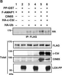
- Western Blot: LOX propeptide Antibody [NBP1-30327] - LOX-PP reduces CIN85 mono-ubiquitination and ability to interact with c-Cbl. HEK293T cells were transfected with AMAP1-FLAG, CIN85, HA-c-Cbl, HA-ubiquitin and LOX-PP-GST (PP-GST) as indicated and subjected to a ubiquitination assay. FLAG-tagged AMAP1 was immunoprecipitated and total whole cell extracts were subjected to WB with the indicated antibodies (lower panel). Data were quantified and relative mono-ubiquitination of AMAP1 with and without LOX-PP was determined by averaging the results of three independent experiments. Image collected and cropped by CiteAb from the following publication (http://dx.plos.org/10.1371/journal.pone.0077288), licensed under a CC-BY licence.

Supportive validation
- Submitted by
- Novus Biologicals (provider)
- Main image

- Experimental details
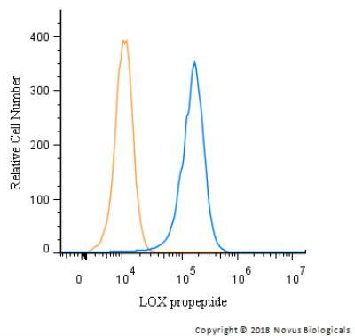
- Flow Cytometry: LOX propeptide Antibody [NBP1-30327] - An intracellular stain was performed on HeLa with LOX propeptide Antibody NBP1-30327 and a matched isotype control. Cells were fixed with 4% PFA and then permeabilized with 0.1% saponin. Cells were incubated in an antibody dilution of 1 ug/mL for 30 minutes at room temperature, followed by Rabbit IgG (H+L) Cross-Adsorbed Secondary Antibody, .