Explore
 Validate
 Learn
Learn Western blot
 Immunocytochemistry
Antibody data
- Antibody Data
- Antigen structure
- References [4]
- Comments [0]
- Validations
- Immunocytochemistry [1]
- Immunohistochemistry [1]
- Other assay [5]
Submit
Validation data
Reference
Comment
Report error
- Product number
- PA1-16953 - Provider product page

- Provider
- Invitrogen Antibodies
- Product name
- LOX Polyclonal Antibody
- Antibody type
- Polyclonal
- Antigen
- Synthetic peptide
- Description
- PA1-16953 is specific for the ~58 kDa LOX protein. This is a highly glycosylated protein and therefore, other bands may be seen at ~32 kDa (non-glycosylated form) 50 kDa and 58 kDa (glycosylated forms). Suggested positive control: human and mouse kidney lysate, antigen standard for LOX (transient overexpression lysate), human kidney protein, mouse kidney protein.
- Reactivity
- Human, Mouse, Rat, Feline, Porcine
- Host
- Rabbit
- Isotype
- IgG
- Vial size
- 100 μL
- Concentration
- 1 mg/mL
- Storage
- -20°C, Avoid Freeze/Thaw Cycles
Submitted references DuCLOX-2/5 Inhibition Attenuates Inflammatory Response and Induces Mitochondrial Apoptosis for Mammary Gland Chemoprevention.
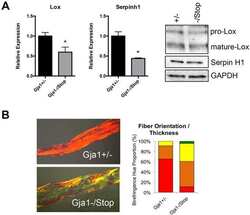
Defective signaling, osteoblastogenesis and bone remodeling in a mouse model of connexin 43 C-terminal truncation.
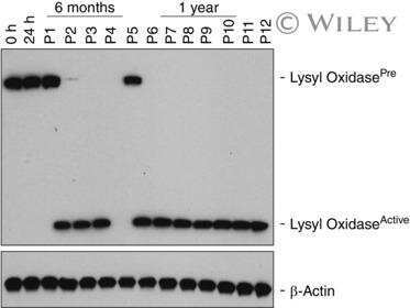
Elevated ischaemia-associated lysyl oxidase activity in delayed graft failure 6-12 months after renal transplantation.
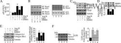
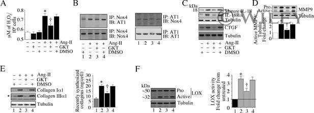
The Nox1/4 Dual Inhibitor GKT137831 or Nox4 Knockdown Inhibits Angiotensin-II-Induced Adult Mouse Cardiac Fibroblast Proliferation and Migration. AT1 Physically Associates With Nox4.
Gautam S, Rawat AK, Sammi SR, Roy S, Singh M, Devi U, Yadav RK, Singh L, Rawat JK, Ansari MN, Saeedan AS, Kumar D, Pandey R, Kaithwas G
Frontiers in pharmacology 2018;9:314
Frontiers in pharmacology 2018;9:314
Defective signaling, osteoblastogenesis and bone remodeling in a mouse model of connexin 43 C-terminal truncation.
Moorer MC, Hebert C, Tomlinson RE, Iyer SR, Chason M, Stains JP
Journal of cell science 2017 Feb 1;130(3):531-540
Journal of cell science 2017 Feb 1;130(3):531-540
Elevated ischaemia-associated lysyl oxidase activity in delayed graft failure 6-12 months after renal transplantation.
Chen Z, Li Y, Xu H, Ma F, Li J, Zhao L, Xu Y
Experimental physiology 2017 Feb 1;102(2):282-287
Experimental physiology 2017 Feb 1;102(2):282-287
The Nox1/4 Dual Inhibitor GKT137831 or Nox4 Knockdown Inhibits Angiotensin-II-Induced Adult Mouse Cardiac Fibroblast Proliferation and Migration. AT1 Physically Associates With Nox4.
Somanna NK, Valente AJ, Krenz M, Fay WP, Delafontaine P, Chandrasekar B
Journal of cellular physiology 2016 May;231(5):1130-41
Journal of cellular physiology 2016 May;231(5):1130-41
No comments: Submit comment
Supportive validation
- Submitted by
- Invitrogen Antibodies (provider)
- Main image
- Experimental details
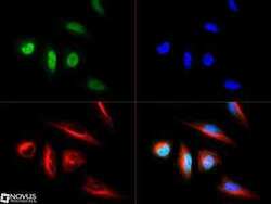
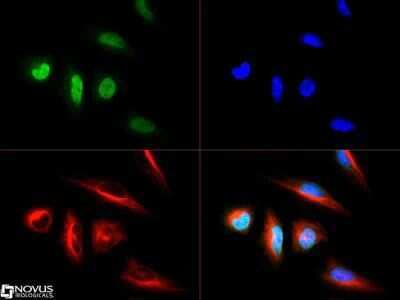
- Immunocytochemistry analysis of LOX in HeLa cells. Samples were incubated in LOX polyclonal antibody (Product # PA1-16953) followed by DyLight 488 (green). Nuclei and alpha-tubulin were counterstained with DAPI (blue) and DyLight 550 (red).
Supportive validation
- Submitted by
- Invitrogen Antibodies (provider)
- Main image
- Experimental details
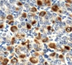
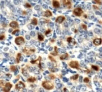
- Immunohistochemical analysis of LOX in mouse stomach. Samples were incubated in LOX polyclonal antibody (Product # PA1-16953).
Supportive validation
- Submitted by
- Invitrogen Antibodies (provider)
- Main image

- Experimental details
- NULL
- Submitted by
- Invitrogen Antibodies (provider)
- Main image
- Experimental details
- NULL
- Submitted by
- Invitrogen Antibodies (provider)
- Main image
- Experimental details
- NULL

- Submitted by
- Invitrogen Antibodies (provider)
- Main image
- Experimental details
- NULL

- Submitted by
- Invitrogen Antibodies (provider)
- Main image

- Experimental details
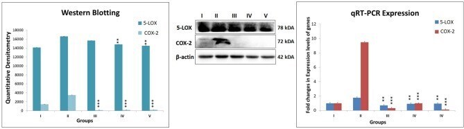
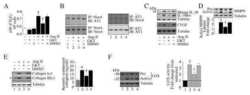
- Figure 6 Expression level of protein of COX-2 and 5-LOX through western blot and levels of gene contributor through quantitative RT-PCR. Immunoblotting of respective individual group [I-Control (Normal saline, 3 ml/kg, p.o.), II- Toxic control (MNU 47 mg/kg, i.v.), III- Zaltoprofen (10 mg/kg, p.o. + MNU 47 mg/kg, i.v.), IV- Zileuton (10 mg/kg, p.o. + MNU 47 mg/kg, i.v.) and V- Zaltoprofen + Zileuton (5 + 5 mg/kg, p.o. + MNU 47 mg/kg, i.v.)] for COX-2 and 5-LOX. Excised mammary gland tissue sample lysed in trizol for RNA extraction and analyzed for the mRNA expression of COX-2 and 5-LOX by qRT-PCR. beta-actin was used as loading control. Each experiment was performed in triplicate. Values are presented as Mean +- SD. Comparisons were made by the one-way ANOVA followed by Bonferroni multiple test. All groups were compared to the MNU treated group ( ** p < 0.01, *** p < 0.001).