 Explore
 Validate
 Learn
Learn Western blot
Antibody data
- Antibody Data
- Antigen structure
- References [3]
- Comments [0]
- Validations
- Western blot [1]
- Immunohistochemistry [1]
Submit
Validation data
Reference
Comment
Report error
- Product number
- PAB10048 - Provider product page

- Provider
- Abnova Corporation
- Proper citation
- Abnova Corporation Cat#PAB10048, RRID:AB_1674715
- Product name
- FANCG polyclonal antibody
- Antibody type
- Polyclonal
- Description
- Rabbit polyclonal antibody raised against synthetic peptide of FANCG.
- Storage
- Store at 4°C. For long term storage store at -20°C.Aliquot to avoid repeated freezing and thawing.
Submitted references The Fanconi anemia core complex associates with chromatin during S phase.
Oxidative stress/damage induces multimerization and interaction of Fanconi anemia proteins.
Telomere dynamics in Fancg-deficient mouse and human cells.
Mi J, Kupfer GM
Blood 2005 Jan 15;105(2):759-66
Blood 2005 Jan 15;105(2):759-66
Oxidative stress/damage induces multimerization and interaction of Fanconi anemia proteins.
Park SJ, Ciccone SL, Beck BD, Hwang B, Freie B, Clapp DW, Lee SH
The Journal of biological chemistry 2004 Jul 16;279(29):30053-9
The Journal of biological chemistry 2004 Jul 16;279(29):30053-9
Telomere dynamics in Fancg-deficient mouse and human cells.
Franco S, van de Vrugt HJ, Fernández P, Aracil M, Arwert F, Blasco MA
Blood 2004 Dec 15;104(13):3927-35
Blood 2004 Dec 15;104(13):3927-35
No comments: Submit comment
Supportive validation
- Submitted by
- Abnova Corporation (provider)
- Main image
- Experimental details
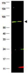
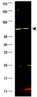
- Western blot using FANCG polyclonal antibody (Cat # PAB10048) shows detection of a band at ~69 KDa (arrowhead) corresponding to FANCG present in a HeLa whole cell lysate.Approximately 35 ug of lysate was separated by 4-20% Tris Glycine SDS-PAGE.After blocking, the membrane was probed overnight at 4°C with the primary antibody diluted to 1:500.The membrane was washed and reacted with a 1:10,000 dilution of IRDye™800 conjugated Gt-a-Rabbit IgG [H&L] for 45 min at room temperature (800 nm channel, green).Molecular weight estimation was made by comparison to prestained MW markers (indicated at left).IRDye™800 fluorescence image was captured using the Odyssey® Infrared Imaging System developed by LI-COR.IRDye is a trademark of LI-COR, Inc.
Supportive validation
- Submitted by
- Abnova Corporation (provider)
- Main image
- Experimental details
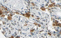
- Immunohistochemistry of FANCG polyclonal antibody (Cat # PAB10048) shows strong nuclear and cytoplasmic staining of cells of macrophages in human lung tissue.Tissue was formalin-fixed and paraffin embedded.Brown color indicates presence of protein, blue color shows cell nuclei.Personal Communication, Kenneth Wester, www.proteinatlas.org, Uppsala, Sweden.
- Validation comment
- Immunohistochemistry (Formalin/PFA-fixed paraffin-embedded sections)
