Explore
 Validate
 Learn
Learn Western blot
 Other assay
Antibody data
- Antibody Data
- Antigen structure
- References [2]
- Comments [0]
- Validations
- Other assay [5]
Submit
Validation data
Reference
Comment
Report error
- Product number
- PA5-46771 - Provider product page

- Provider
- Invitrogen Antibodies
- Product name
- PDZD8 Polyclonal Antibody
- Antibody type
- Polyclonal
- Antigen
- Synthetic peptide
- Description
- Peptide sequence: TRHIINTSSR LLNLRQVSKT RLSEPGTDLV EPSPKHTPNT SDNEGSDTEV
- Reactivity
- Human
- Host
- Rabbit
- Isotype
- IgG
- Vial size
- 100 μL
- Concentration
- 0.5 mg/mL
- Storage
- -20°C, Avoid Freeze/Thaw Cycles
Submitted references circ_0020123 promotes cell proliferation and migration in lung adenocarcinoma via PDZD8.
MERLIN: a novel BRET-based proximity biosensor for studying mitochondria-ER contact sites.
Wei W, Wang C, Wang L, Zhang J
Open medicine (Warsaw, Poland) 2022;17(1):536-549
Open medicine (Warsaw, Poland) 2022;17(1):536-549
MERLIN: a novel BRET-based proximity biosensor for studying mitochondria-ER contact sites.
Hertlein V, Flores-Romero H, Das KK, Fischer S, Heunemann M, Calleja-Felipe M, Knafo S, Hipp K, Harter K, Fitzgerald JC, García-Sáez AJ
Life science alliance 2020 Jan;3(1)
Life science alliance 2020 Jan;3(1)
No comments: Submit comment
Supportive validation
- Submitted by
- Invitrogen Antibodies (provider)
- Main image
- Experimental details
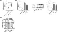
- Figure 1 PDZD8 is highly expressed in LUAD tissues and cells. (a) PDZD8 expression in LUAD tissues ( n = 16) was elevated compared with that in normal tissues ( n = 16) as shown by RT-qPCR. ** p < 0.01. (b) The mRNA and protein levels of PDZD8 in LUAD cells were increased as suggested by RT-qPCR and western blot analyses. ** p < 0.01, *** p < 0.001 vs BEAS-2B group. (c) Subcellular fractionation assays revealed that PDZD8 was mainly distributed in cytoplasm of LUAD cells. The data are presented as the mean value +- standard deviation. Student's t test was used to compare differences between LUAD tissues and normal tissues. One-way analysis of variance followed by Tukey's post hoc analysis was used to compare differences among groups.

- Submitted by
- Invitrogen Antibodies (provider)
- Main image

- Experimental details
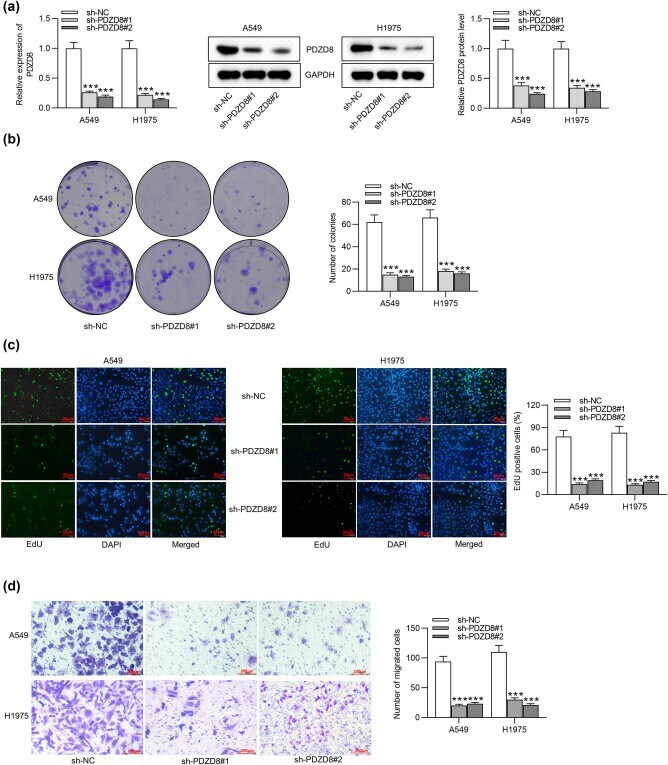
- Figure 2 PDZD8 knockdown inhibits proliferation and migration of LUAD cells. (a) PDZD8 mRNA expression and protein levels were decreased by sh-PDZD8#1/2 in A549 and H1975 cells according to RT-qPCR and western blot. (b and c) Colony formation assays and EdU assays suggested the suppressive effect of PDZD8 knockdown on LUAD cell proliferation. (d and e) Transwell and wound healing assays revealed that PDZD8 silencing inhibited the migration of LUAD cells. The data are presented as the mean value +- standard deviation. One-way analysis of variance followed by Tukey's post hoc analysis was used to compare differences among groups. *** p < 0.001 vs sh-NC group.
- Submitted by
- Invitrogen Antibodies (provider)
- Main image
- Experimental details
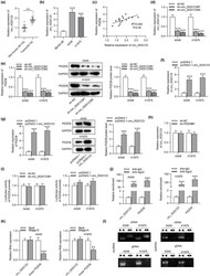
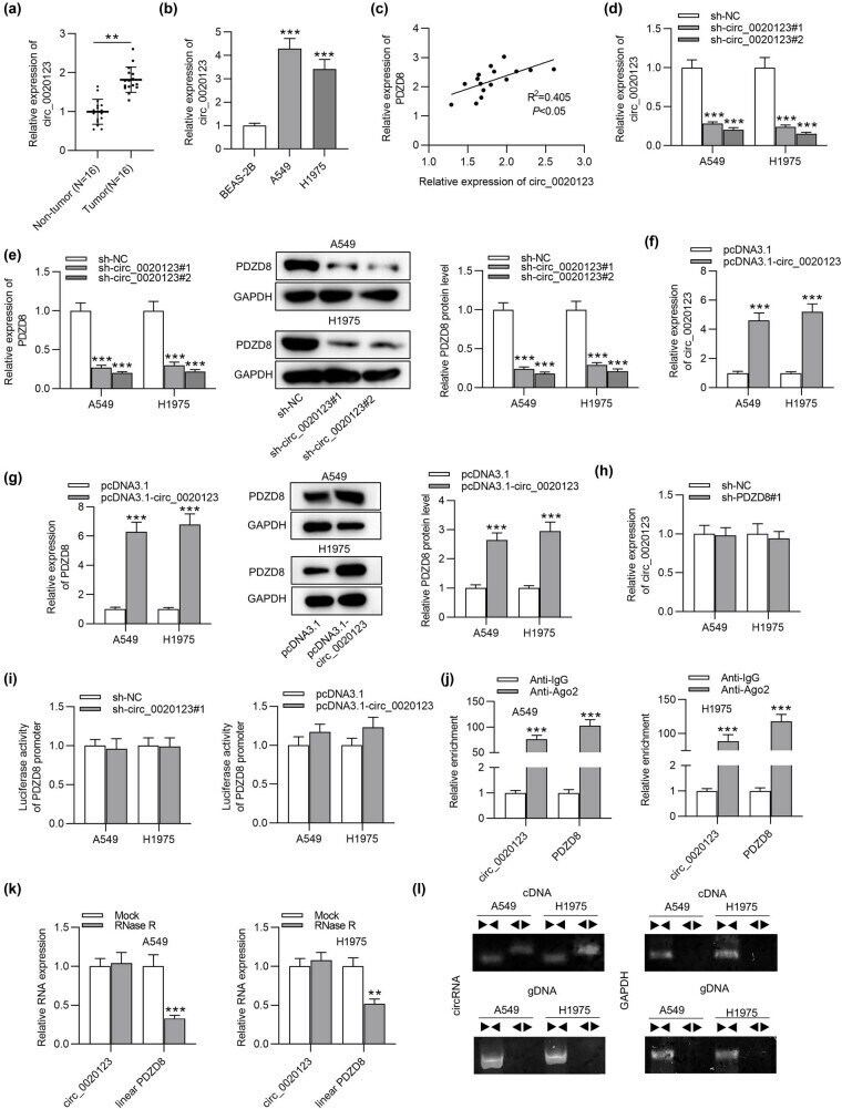
- Figure 3 circ_0020123 positively regulates PDZD8 expression in LUAD tissues and cells. (a) circ_0020123 expression was upregulated in LUAD tissues as detected by RT-qPCR. ** p < 0.01. (b) circ_0020123 showed high expression in LUAD cells as shown by RT-qPCR. *** p < 0.001 vs BEAS-2B group. (c) A positive expression correlation between circ_0020123 and PDZD8 in LUAD tissues was identified using Spearman correlation coefficient. R 2 = 0.405, * p < 0.05. (d) circ_0020123 expression was successfully knocked down in LUAD cells as shown by RT-qPCR. *** p < 0.001 vs sh-NC group. (e) circ_0020123 knockdown reduced PDZD8 expression at both the mRNA and protein levels. *** p < 0.001 vs sh-NC group. (f) RT-qPCR was performed to detect the efficiency of circ_0020123 overexpression. *** p < 0.001 vs pcDNA3.1 group. (g) The influence of overexpressed circ_0020123 on PDZD8 mRNA expression and protein levels in LUAD cells was quantified by RT-qPCR and western blot analyses. *** p < 0.001 vs pcDNA3.1 group. (h) The effect of PDZD8 knockdown on circ_0020123 expression in LUAD cells were examined by RT-qPCR. (i) Luciferase reporter assays were conducted to explore the effect of circ_0020123 on the transcription level of PDZD8. (j) RIP assays implied that circ_0020123 and PDZD8 were enriched in RNA-induced silencing complex. *** p < 0.001 vs Anti-IgG group. (k) The expression levels of circ_0020123 and linear PDZD8 in A549 and H1975 cells after RNase R treatment were determined by RT-qPCR analys
- Submitted by
- Invitrogen Antibodies (provider)
- Main image
- Experimental details
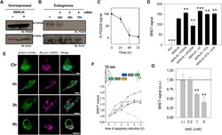
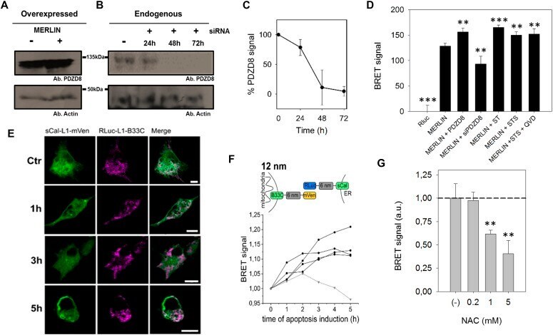
- Figure 3. Validation of MERLIN. (A, B, C, D) PDZD8 modulates ER-mitochondria distance. (A, B, C) Representative Western blot of the PDZD8 levels when transiently transfected and (B) upon silencing with siRNA_PDZD8 in HCT116 cells, whose quantification is shown in (C) (n = 3). (D) BRET signal in cells co-expressing Rluc-L1-B33C and Scal-L1-mVenus biosensor combination, in the presence of overexpressed PDZD8, the synthetic tether mTagBFP2 and PDZD8 knockdown in HCT116 cells. (** P < 0.025, *** P
- Submitted by
- Invitrogen Antibodies (provider)
- Main image
- Experimental details
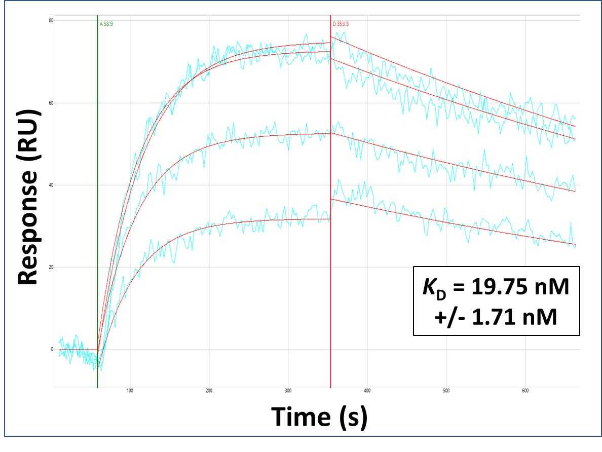
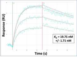
- Surface Plasmon Resonance of PDZD8 polyclonal antibody (Product # PA5-46771). Purified polyclonal antibodies were immobilized on a Protein A/G coated Carterra LSA sensor chip at concentrations of 5, and 50 µg/mL in duplicate. Antibodies on the surface were exposed to interaction with peptides sequentially via microfluidic controlled flow at 333 nm peptide concentration for kinetic characterization of the binders for affinity and specificity, followed by curve fitting using the Kinetics software. Kd determinations for both concentrations were averaged and results and standard deviation are shown.