Explore
 Validate
 Learn
Learn Western blot
 ELISA
ELISAAntibody data
- Antibody Data
- Antigen structure
- References [0]
- Comments [0]
- Validations
- Western blot [2]
- Immunohistochemistry [1]
- Flow cytometry [1]
Submit
Validation data
Reference
Comment
Report error
- Product number
- OAAB03615 - Provider product page

- Provider
- Aviva Systems Biology
- Product name
- WNT10B antibody - center region
- Antibody type
- Polyclonal
- Reactivity
- Human, Mouse
- Host
- Rabbit
- Vial size
- 400ul
- Storage
- Maintain refrigerated at 2-8 deg C for up to 6 months. For long term storage store at -20 deg C in small aliquots to prevent freeze-thaw cycles.
No comments: Submit comment
Supportive validation
- Submitted by
- Aviva Systems Biology (provider)
- Main image
- Experimental details
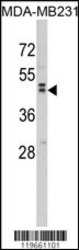
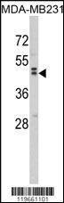
- Western blot analysis of WNT10B Antibody (Center) in MDA-MB231 cell lysates (35ug/lane). WNT10B (arrow) was detected using the purified Pab.
- Sample type
- MDA-MB231 cell line lysates
- Primary Ab dilution
- 1.0 µg/mL
- Protocol
- Protocol
- Submitted by
- Aviva Systems Biology (provider)
- Main image
- Experimental details
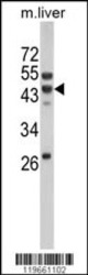
- Western blot analysis of WNT10B Antibody (Center) in mouse liver tissue lysates (35ug/lane). WNT10B (arrow) was detected using the purified Pab.
- Sample type
- mouse liver tissue lysates
- Primary Ab dilution
- 1.0 µg/mL
- Protocol
- Protocol

Supportive validation
- Submitted by
- Aviva Systems Biology (provider)
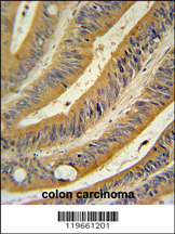
- Main image

- Experimental details
- WNT10B Antibody (Center) immunohistochemistry analysis in formalin fixed and paraffin embedded human colon carcinoma followed by peroxidase conjugation of the secondary antibody and DAB staining. This data demonstrates the use of the WNT10B Antibody (Center) for immunohistochemistry. Clinical relevance has not been evaluated.
- Sample type
- human colon carcinoma
- Primary Ab dilution
- 1.0 µg/mL
- Protocol
- Protocol
Supportive validation
- Submitted by
- Aviva Systems Biology (provider)
- Main image

- Experimental details
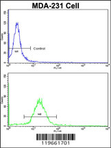
- Flow cytometric analysis of MDA-231 cells using WNT10B Antibody (Center)(bottom histogram) compared to a negative control cell (top histogram). FITC-conjugated goat-anti-rabbit secondary antibodies were used for the analysis.
- Sample type
- MDA-231 cells
- Primary Ab dilution
- 1.0 µg/mL
- Protocol
- Protocol