Explore
 Validate
 Learn
Learn Western blot
Antibody data
- Antibody Data
- Antigen structure
- References [0]
- Comments [0]
- Validations
- Western blot [1]
- Immunohistochemistry [7]
Submit
Validation data
Reference
Comment
Report error
- Product number
- NB100-56567 - Provider product page

- Provider
- Novus Biologicals
- Proper citation
- Novus Cat#NB100-56567, RRID:AB_2058238
- Product name
- Mouse Monoclonal APIP Antibody
- Antibody type
- Monoclonal
- Description
- Protein G purified. This monoclonal antibody can be used for detection of APIP and APIP2.
- Reactivity
- Human
- Host
- Mouse
- Isotype
- IgG
- Vial size
- 0.1 mg
- Concentration
- 0.5 mg/ml
- Storage
- Store at 4C short term. Aliquot and store at -20C long term. Avoid freeze-thaw cycles.
No comments: Submit comment
Supportive validation
- Submitted by
- Novus Biologicals (provider)
- Main image
- Experimental details
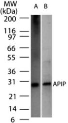
- Western Blot: APIP Antibody (19F461) [NB100-56567] - Analysis of APIP2 in (A) recombinant protein and (B) HeLa cell lysate using APIP antibody at 2 ug/ml.

Supportive validation
- Submitted by
- Novus Biologicals (provider)
- Main image
- Experimental details
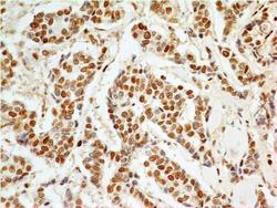
- Immunohistochemistry-Paraffin: APIP Antibody (19F461) [NB100-56567] - Formalin-fixed, paraffin-embedded adenocarcinoma of the breast stained with APIP antibody (5 ug/ml), peroxidase-conjugate and DAB chromogen. Staining of formalin-fixed tissues is enhanced by boiling tissue sections in 10 mM sodium citrate buffer, pH 6.0 for 10-20 min followed by cooling at RT for 20 min.

- Submitted by
- Novus Biologicals (provider)
- Main image
- Experimental details
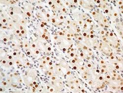
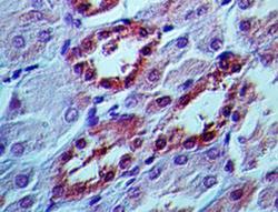
- Immunohistochemistry-Paraffin: APIP Antibody (19F461) [NB100-56567] - Formalin-fixed, paraffin-embedded human stomach stained with APIP antibody (5 ug/ml), peroxidase-conjugate and DAB chromogen. Staining of formalin-fixed tissues is enhanced by boiling tissue sections in 10 mM sodium citrate buffer, pH 6.0 for 10-20 min followed by cooling at RT for 20 min.
- Submitted by
- Novus Biologicals (provider)
- Main image
- Experimental details
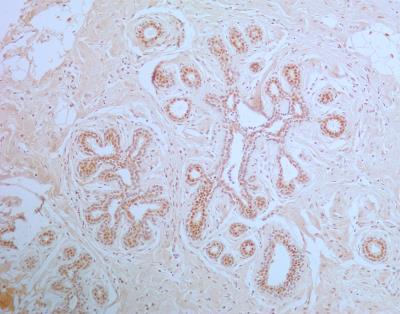
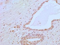
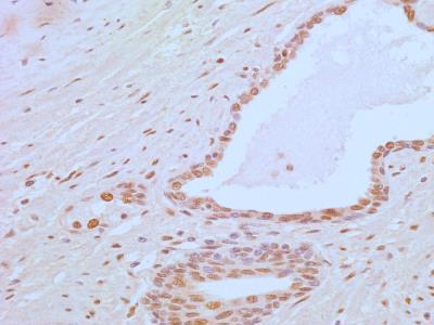
- Immunohistochemistry-Paraffin: APIP Antibody (19F461) [NB100-56567] - Analysis of APIP in a section of normal prostate from human using 5 ug/ml concentration of APIP antibody (clone 19F461). The tubuloalveolar glands in prostate section depicted APIP positivity in the cytoplasm and nuclei of the epithelial cells. The fibro-muscular stroma showed an overall weak staining with intense nuclear positivity in some cells.
- Submitted by
- Novus Biologicals (provider)
- Main image
- Experimental details
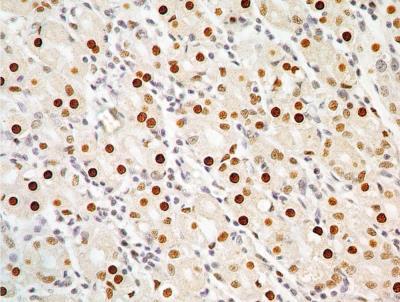
- Immunohistochemistry-Paraffin: APIP Antibody (19F461) [NB100-56567] - Analysis of formalin-fixed paraffin-embedded human kidney tissue using APIP antibody at 5 ug/ml concentration.

- Submitted by
- Novus Biologicals (provider)
- Main image
- Experimental details
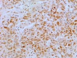
- Immunohistochemistry-Paraffin: APIP Antibody (19F461) [NB100-56567] - Analysis of human lung cancer tissue section using APIP antibody (clone 19F461) at a concentration of 5 ug/ml. The representative image shows a nuclear and cytoplasmic staining pattern of APIP expression in lung cancer cells.

- Submitted by
- Novus Biologicals (provider)
- Main image
- Experimental details
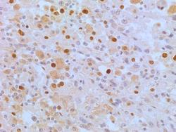
- Immunohistochemistry-Paraffin: APIP Antibody (19F461) [NB100-56567] - Analysis of human renal cancer tissue section using APIP antibody (clone 19F461) at a concentration of 5 ug/ml. The representative image shows a nuclear and cytoplasmic staining pattern of APIP expression in cancer cells.
- Submitted by
- Novus Biologicals (provider)
- Main image

- Experimental details
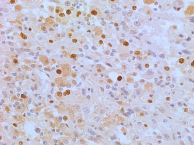
- Immunohistochemistry-Paraffin: APIP Antibody (19F461) [NB100-56567] - Detection of APIP in a section of normal human breast tissue using APIP antibody (clone 19F461) at a concentration of 5 ug/ml. The breast's ductal/acinar epithelial cells showed strong cytoplasmic as well as nuclear expression, whereas the myoepithelial cells and the intra-lobular connective tissue depicted very weak to negligible APIP positivity.