 Explore
 Validate
 Learn
Learn Western blot
Antibody data
- Antibody Data
- Antigen structure
- References [3]
- Comments [0]
- Validations
- Western blot [1]
- Immunocytochemistry [2]
- Immunohistochemistry [3]
- Flow cytometry [1]
Submit
Validation data
Reference
Comment
Report error
- Product number
- A21371 - Provider product page

- Provider
- Invitrogen Antibodies
- Product name
- alpha Tubulin Monoclonal Antibody (236-10501), Biotin
- Antibody type
- Monoclonal
- Antigen
- Other
- Description
- Storage and reconstitution: upon receipt, store desiccated at -20ºC. To prepare a stock solution, reconstitute in PBS containing 1% BSA and 2mM sodium azide. Reconstituted solutions should remain stable for 6 months when stored at 4ºC. For longer storage, aliquot and store at -20ºC.
- Reactivity
- Human, Mouse, Bovine
- Host
- Mouse
- Conjugate
- Biotin
- Isotype
- IgG
- Antibody clone number
- 236-10501
- Vial size
- 50 µg
- Storage
- -20°C
Submitted references Mapping the 3D orientation of piconewton integrin traction forces.
Glycolytic requirement for NK cell cytotoxicity and cytomegalovirus control.
A single class II myosin modulates T cell motility and stopping, but not synapse formation.
Brockman JM, Blanchard AT, Pui-Yan V Ma, Derricotte WD, Zhang Y, Fay ME, Lam WA, Evangelista FA, Mattheyses AL, Salaita K
Nature methods 2018 Feb;15(2):115-118
Nature methods 2018 Feb;15(2):115-118
Glycolytic requirement for NK cell cytotoxicity and cytomegalovirus control.
Mah AY, Rashidi A, Keppel MP, Saucier N, Moore EK, Alinger JB, Tripathy SK, Agarwal SK, Jeng EK, Wong HC, Miller JS, Fehniger TA, Mace EM, French AR, Cooper MA
JCI insight 2017 Dec 7;2(23)
JCI insight 2017 Dec 7;2(23)
A single class II myosin modulates T cell motility and stopping, but not synapse formation.
Jacobelli J, Chmura SA, Buxton DB, Davis MM, Krummel MF
Nature immunology 2004 May;5(5):531-8
Nature immunology 2004 May;5(5):531-8
No comments: Submit comment
Supportive validation
- Submitted by
- Invitrogen Antibodies (provider)
- Main image

- Experimental details
- Western blot analysis of alpha-Tubulin was performed by loading 20 µg of HCT 116 (lane1), MDA-MB-231 (lane2), Raji (lane3) and A431 (lane4) cell lysate using Novex®NuPAGE® 12 % Bis-Tris gel (Product # NP0341BOX), XCell SureLock Electrophoresis System (Product # EI0002), Novex® Sharp Pre-Stained Protein Standard (LC5800), and iBlot® Dry Blotting System (IB21001). Proteins were transferred to a nitrocellulose membrane and blocked with 5% skim milk at 4°C overnight. alpha-Tubulin was detected at ~ 52 kDa using alpha-Tubulin Biotin Conjugated Mouse Monoclonal Antibody (Product # A21371) at 1:3000 dilution in 5% skim milk for 3 hour at room temperature on a rocking platform. Streptavidin - HRP (Product # SA1007) at 1:2000 dilution was used and chemiluminescent detection was performed using Pierce™ ECL Western Blotting Substrate (Product # 32106).
- Conjugate
- Biotin

Supportive validation
- Submitted by
- Invitrogen Antibodies (provider)
- Main image

- Experimental details
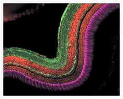
- Binucleate bovine pulmonary artery endothelial cell. Anti-α-tubulin, biotin-XX conjugate; Alexa Fluor® 568 streptavidin and nuclear yellow. A binucleate bovine pulmonary artery endothelial cell labeled with the biotin-XX conjugate of anti-α-tubulin antibody (Product # A21371) and Alexa Fluor® 568 streptavidin (Product # S-11226), then counterstained with nuclear yellow (Product # N21485).
- Conjugate
- Biotin

- Submitted by
- Invitrogen Antibodies (provider)
- Main image
- Experimental details
- Multinucleate HeLa cell in metaphase: DAPI, anti-bovine α-tubulin, mouse monoclonal 236-10501, biotin-XX conjugate and Alexa Fluor® 568 streptavidin. A multinucleate HeLa cell in metaphase that was fixed and then stained with a combination of fluorescent dyes. The chromosomes were stained with DAPI (Product # D1306, D3571, D21490). The cytoskeleton was detected with the biotin-XX conjugate of mouse monoclonal anti-α-tubulin antibody (Product # A21371), which was then visualized with red-fluorescent Alexa Fluor® 568 streptavidin (Product # S-11226). The multiple-exposure image was acquired using filter sets appropriate for rhodamine and DAPI.
- Conjugate
- Biotin

Supportive validation
- Submitted by
- Invitrogen Antibodies (provider)
- Main image
- Experimental details
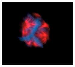
- Anti-bovine α-tubulin, biotin-XX conjugate; Alexa Fluor 647 goat anti-mouse IgG (H+L) conjugate; SYTOX Orange nucleic acid stain. A zebrafish cryosection incubated with the biotin-XX conjugate of mouse monoclonal anti-α-tubulin antibody (Product # A21371). The signal was amplified with TSA Kit #22, which includes HRP-streptavidin and Alexa Fluor 488 tyramide (Product # T-20932). The sample was then incubated with the mouse monoclonal FRet 6 antibody and was visualized with Alexa Fluor 647 goat anti-mouse IgG (Product # A-21235), which is pseudocolored magenta. Finally, the nuclei were counterstained with SYTOX Orange nucleic acid stain (Product # S11368).
- Conjugate
- Biotin

- Submitted by
- Invitrogen Antibodies (provider)
- Main image
- Experimental details
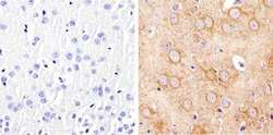
- Immunohistochemistry analysis of Alpha-Tubulin showing staining in the cytoplasm of paraffin-embedded mouse brain tissue (right) compared to a negative control without primary antibody (left). To expose target proteins, antigen retrieval was performed using 10mM sodium citrate (pH 6.0), microwaved for 8-15 min. Following antigen retrieval, tissues were blocked in 3% H2O2-methanol for 15 min at room temperature, washed with ddH2O and PBS, and then probed with a Alpha-Tubulin (Product # A21371) diluted in 3% BSA-PBS at a dilution of 1:20 overnight at 4°C in a humidified chamber. Tissues were washed extensively in PBST and detection was performed using an HRP-conjugated secondary antibody followed by colorimetric detection using a DAB kit. Tissues were counterstained with hematoxylin and dehydrated with ethanol and xylene to prep for mounting.
- Conjugate
- Biotin

- Submitted by
- Invitrogen Antibodies (provider)
- Main image

- Experimental details
- Immunohistochemistry analysis of Alpha-Tubulin showing staining in the cytoplasm of paraffin-embedded human breast carcinoma (right) compared to a negative control without primary antibody (left). To expose target proteins, antigen retrieval was performed using 10mM sodium citrate (pH 6.0), microwaved for 8-15 min. Following antigen retrieval, tissues were blocked in 3% H2O2-methanol for 15 min at room temperature, washed with ddH2O and PBS, and then probed with a Alpha-Tubulin (Product # A21371) diluted in 3% BSA-PBS at a dilution of 1:20 overnight at 4°C in a humidified chamber. Tissues were washed extensively in PBST and detection was performed using an HRP-conjugated secondary antibody followed by colorimetric detection using a DAB kit. Tissues were counterstained with hematoxylin and dehydrated with ethanol and xylene to prep for mounting.
- Conjugate
- Biotin

Supportive validation
- Submitted by
- Invitrogen Antibodies (provider)
- Main image
- Experimental details
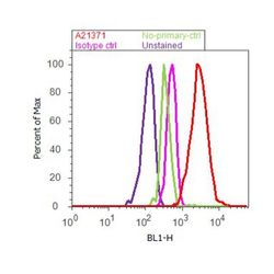
- Flow cytometry analysis of alpha-Tubulin was done on HeLa cells. Cells were fixed with 70% ethanol for 10 minutes, permeabilized with 0.25% Tritonª X-100 for 20 minutes, and blocked with 5% BSA for 30 minutes at room temperature. Cells were labeled with alpha-Tubulin Biotin Conjugated Mouse Monoclonal Antibody (A21371, red histogram) or with mouse isotype control (pink histogram) at 3-5 µg/million cells in 2.5% BSA. After incubation at room temperature for 2 hours, the cells were labeled with Biotin conjugated Goat Anti-Mouse Secondary Antibody (A16082) followed by Streptavidin, Alexa Fluor¨ 488 conjugate (S-11223) at a dilution of 1:400 each for 30 minutes at room temperature. The representative 10,000 cells were acquired and analyzed for each sample using an Attune¨ Acoustic Focusing Cytometer. The purple histogram represents unstained control cells and the green histogram represents no-primary-antibody control.
