Explore
 Validate
 Learn
Learn Western blot
Antibody data
- Antibody Data
- Antigen structure
- References [2]
- Comments [0]
- Validations
- Western blot [2]
- Immunocytochemistry [1]
- Immunohistochemistry [1]
Submit
Validation data
Reference
Comment
Report error
- Product number
- AF6120 - Provider product page

- Provider
- R&D Systems
- Product name
- Mouse/Rat Semaphorin 4C Antibody
- Antibody type
- Polyclonal
- Description
- Antigen Affinity-purified. Detects mouse and rat Semaphorin 4C in Western blots. In direct ELISAs, less than 20% cross-reactivity with recombinant human Semaphorin 4C and less than 5% cross-reactivity with recombinant mouse (rm) Semaphorin 4B, rmSemaphorin 4D, and rmSemaphorin 4G is observed.
- Reactivity
- Mouse, Rat
- Host
- Sheep
- Conjugate
- Unconjugated
- Antigen sequence
Q64151- Isotype
- IgG
- Vial size
- 100 ug
- Concentration
- LYOPH
- Storage
- Use a manual defrost freezer and avoid repeated freeze-thaw cycles. 12 months from date of receipt, -20 to -70 °C as supplied. 1 month, 2 to 8 °C under sterile conditions after reconstitution. 6 months, -20 to -70 °C under sterile conditions after reconstitution.
Submitted references Plexin B2 and Semaphorin 4C Guide T Cell Recruitment and Function in the Germinal Center.
Semaphorin 4C Protects against Allergic Inflammation: Requirement of Regulatory CD138+ Plasma Cells.
Yan H, Wu L, Shih C, Hou S, Shi J, Mao T, Chen W, Melvin B, Rigby RJ, Chen Y, Jiang H, Friedel RH, Vinuesa CG, Qi H
Cell reports 2017 May 2;19(5):995-1007
Cell reports 2017 May 2;19(5):995-1007
Semaphorin 4C Protects against Allergic Inflammation: Requirement of Regulatory CD138+ Plasma Cells.
Xue D, Kaufman GN, Dembele M, Beland M, Massoud AH, Mindt BC, Fiter R, Fixman ED, Martin JG, Friedel RH, Divangahi M, Fritz JH, Mazer BD
Journal of immunology (Baltimore, Md. : 1950) 2017 Jan 1;198(1):71-81
Journal of immunology (Baltimore, Md. : 1950) 2017 Jan 1;198(1):71-81
No comments: Submit comment
Supportive validation
- Submitted by
- R&D Systems (provider)
- Main image

- Experimental details
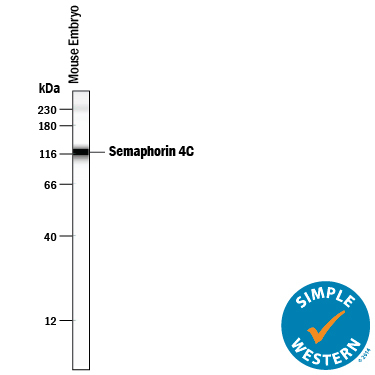
- Detection of Mouse and Rat Semaphorin 4C by Simple WesternTM. Simple Western lane view shows lysates of mouse embryo tissue (15 d.p.c.), loaded at 0.2 mg/mL. A specific band was detected for Semaphorin 4C at approximately 117 kDa (as indicated) using 50 µg/mL of Sheep Anti-Mouse/Rat Semaphorin 4C Antigen Affinity-purified Polyclonal Antibody (Catalog # AF6120) followed by 1:50 dilution of HRP-conjugated Anti-Sheep IgG Secondary Antibody (Catalog # HAF016). This experiment was conducted under reducing conditions and using the 12-230 kDa separation system. Non-specific interaction with the 230 kDa Simple Western standard may be seen with this antibody.
- Submitted by
- R&D Systems (provider)
- Main image
- Experimental details
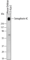
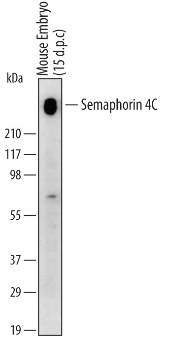
- Detection of Mouse Semaphorin 4C by Western Blot (Non-Reducing). Western blot shows lysates of mouse embryo (15 d.p.c.) tissue. PVDF membrane was probed with 1 µg/mL of Sheep Anti-Mouse/Rat Semaphorin 4C Antigen Affinity-purified Polyclonal Antibody (Catalog # AF6120) followed by HRP-conjugated Anti-Sheep IgG Secondary Antibody (Catalog # HAF016). A specific band was detected for Semaphorin 4C at approximately 240 kDa (as indicated). This experiment was conducted under non-reducing conditions and using Immunoblot Buffer Group 1.
Supportive validation
- Submitted by
- R&D Systems (provider)
- Main image
- Experimental details
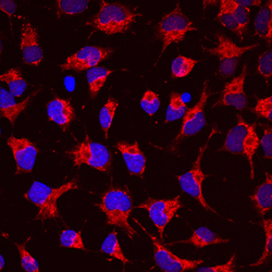
- Semaphorin 4C in Rat Cortical Stem Cells. Semaphorin 4C was detected in immersion fixed rat cortical stem cells using Sheep Anti-Mouse/Rat Semaphorin 4C Antigen Affinity-purified Polyclonal Antibody (Catalog # AF6120) at 10 µg/mL for 3 hours at room temperature. Cells were stained using the NorthernLights™ 557-conjugated Anti-Sheep IgG Secondary Antibody (red; Catalog # NL010) and counterstained with DAPI (blue). Specific staining was localized to cytoplasm. View our protocol for Fluorescent ICC Staining of Cells on Coverslips.

Supportive validation
- Submitted by
- R&D Systems (provider)
- Main image

- Experimental details
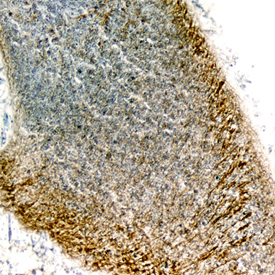
- Semaphorin 4C in Mouse Embryo. Semaphorin 4C was detected in perfusion fixed frozen sections of mouse embryo (13 d.p.c.) using Sheep Anti-Mouse/Rat Semaphorin 4C Antigen Affinity-purified Polyclonal Antibody (Catalog # AF6120) at 1.7 µg/mL overnight at 4 °C. Tissue was stained using the Anti-Sheep HRP-DAB Cell & Tissue Staining Kit (brown; Catalog # CTS019) and counterstained with hematoxylin (blue). Specific staining was localized to neuronal processes of the developing cerebellum. View our protocol for Chromogenic IHC Staining of Frozen Tissue Sections.