 Explore
 Validate
 Learn
Learn Western blot
Antibody data
- Antibody Data
- Antigen structure
- References [1]
- Comments [0]
- Validations
- Western blot [2]
- Immunohistochemistry [1]
Submit
Validation data
Reference
Comment
Report error
- Product number
- AF6125 - Provider product page

- Provider
- R&D Systems
- Product name
- Human Semaphorin 4C Antibody
- Antibody type
- Polyclonal
- Description
- Antigen Affinity-purified. Detects human Semaphorin 4C in direct ELISAs and Western blots. In direct ELISAs, less than 15% cross-reactivity with recombinant mouse (rm) Sema4C is observed and less than 5% cross-reactivity with recombinant human (rh) Sema4G, rhSema4D, rhSema4B and rhSema4A is observed.
- Reactivity
- Human
- Host
- Sheep
- Conjugate
- Unconjugated
- Antigen sequence
Q9C0C4- Isotype
- IgG
- Vial size
- 100 ug
- Concentration
- LYOPH
- Storage
- Use a manual defrost freezer and avoid repeated freeze-thaw cycles. 12 months from date of receipt, -20 to -70 °C as supplied. 1 month, 2 to 8 °C under sterile conditions after reconstitution. 6 months, -20 to -70 °C under sterile conditions after reconstitution.
Submitted references A systems biology approach to characterize the regulatory networks leading to trabectedin resistance in an in vitro model of myxoid liposarcoma.
Uboldi S, Calura E, Beltrame L, Fuso Nerini I, Marchini S, Cavalieri D, Erba E, Chiorino G, Ostano P, D'Angelo D, D'Incalci M, Romualdi C
PloS one 2012;7(4):e35423
PloS one 2012;7(4):e35423
No comments: Submit comment
Supportive validation
- Submitted by
- R&D Systems (provider)
- Main image
- Experimental details
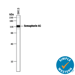
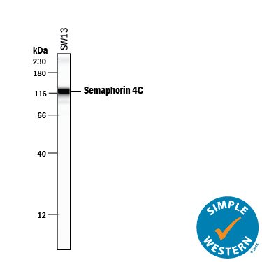
- Detection of Human Semaphorin 4C by Simple WesternTM. Simple Western lane view shows lysates of SW13 human adrenal cortex adenocarcinoma cell line, loaded at 0.2 mg/mL. A specific band was detected for Semaphorin 4C at approximately 121 kDa (as indicated) using 25 µg/mL of Sheep Anti-Human Semaphorin 4C Antigen Affinity-purified Polyclonal Antibody (Catalog # AF6125) followed by 1:50 dilution of HRP-conjugated Anti-Sheep IgG Secondary Antibody (Catalog # HAF016). This experiment was conducted under reducing conditions and using the 12-230 kDa separation system. Non-specific interaction with the 230 kDa Simple Western standard may be seen with this antibody.
- Submitted by
- R&D Systems (provider)
- Main image
- Experimental details
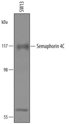
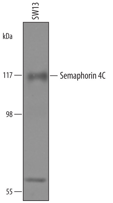
- Detection of Human Semaphorin 4C by Western Blot. Western blot shows lysates of SW13 human adrenal cortex adenocarcinoma cell line. PVDF membrane was probed with 0.5 µg/mL of Sheep Anti-Human Semaphorin 4C Antigen Affinity-purified Polyclonal Antibody (Catalog # AF6125) followed by HRP-conjugated Anti-Sheep IgG Secondary Antibody (Catalog # HAF016). A specific band was detected for Semaphorin 4C at approximately 110 kDa (as indicated). This experiment was conducted under reducing conditions and using Immunoblot Buffer Group 8.
Supportive validation
- Submitted by
- R&D Systems (provider)
- Main image
- Experimental details
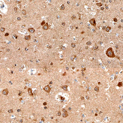
- Semaphorin 4C in Human Brain. Semaphorin 4C was detected in immersion fixed paraffin-embedded sections of human brain (cortex) using Sheep Anti-Human Semaphorin 4C Antigen Affinity-purified Polyclonal Antibody (Catalog # AF6125) at 15 µg/mL overnight at 4 °C. Before incubation with the primary antibody, tissue was subjected to heat-induced epitope retrieval using Antigen Retrieval Reagent-Basic (Catalog # CTS013). Tissue was stained using the Anti-Sheep HRP-DAB Cell & Tissue Staining Kit (brown; Catalog # CTS019) and counterstained with hematoxylin (blue). Specific staining was localized to neuronal cell bodies. View our protocol for Chromogenic IHC Staining of Paraffin-embedded Tissue Sections.
