 Explore
 Validate
 Learn
Learn Western blot
 Immunocytochemistry
Antibody data
- Antibody Data
- Antigen structure
- References [0]
- Comments [0]
- Validations
- Immunocytochemistry [5]
- Immunoprecipitation [1]
- Immunohistochemistry [1]
- Other assay [1]
Submit
Validation data
Reference
Comment
Report error
- Product number
- PA5-21339 - Provider product page

- Provider
- Invitrogen Antibodies
- Product name
- SEC13 Polyclonal Antibody
- Antibody type
- Polyclonal
- Antigen
- Recombinant full-length protein
- Description
- Recommended positive controls: 293T, A431, H1299, HeLa, HepG2, Molt-4, Raji. Predicted reactivity: Mouse (97%), Rat (97%), Zebrafish (87%), Xenopus laevis (88%), Chicken (94%), Rhesus Monkey (98%), Chimpanzee (99%), Bovine (96%). Store product as a concentrated solution. Centrifuge briefly prior to opening the vial.
- Reactivity
- Human
- Host
- Rabbit
- Isotype
- IgG
- Vial size
- 100 μL
- Concentration
- 1 mg/mL
- Storage
- Store at 4°C short term. For long term storage, store at -20°C, avoiding freeze/thaw cycles.
No comments: Submit comment
Supportive validation
- Submitted by
- Invitrogen Antibodies (provider)
- Main image
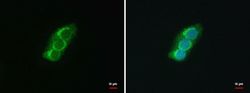
- Experimental details
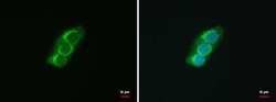
- Immunofluorescent analysis of SEC13 protein isoform 1 showing staining in the vesicles of HepG2 cells. HepG2 cells were fixed in 4% paraformaldehyde at RT for 15 min and stained using a SEC13 protein isoform 1 polyclonal antibody (Product # PA5-21339) diluted at 1:500. Blue: Hoechst 33342 staining.
- Submitted by
- Invitrogen Antibodies (provider)
- Main image
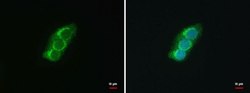
- Experimental details
- Immunofluorescent analysis of SEC13 protein isoform 1 showing staining in the vesicles of HepG2 cells. HepG2 cells were fixed in 4% paraformaldehyde at RT for 15 min and stained using a SEC13 protein isoform 1 polyclonal antibody (Product # PA5-21339) diluted at 1:500. Blue: Hoechst 33342 staining.
- Submitted by
- Invitrogen Antibodies (provider)
- Main image
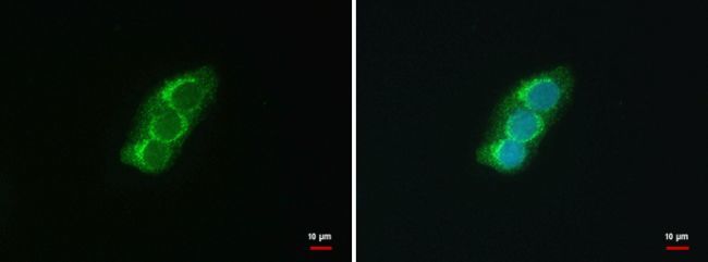
- Experimental details
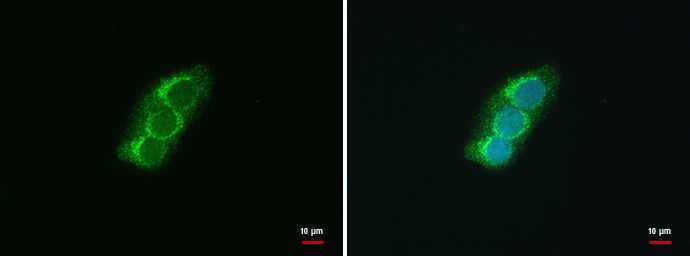
- SEC13 Polyclonal Antibody detects SEC13L1 protein at COP II vesicles by immunofluorescent analysis. Sample: HepG2 cells were fixed in 4% paraformaldehyde at RT for 15 min. Green: SEC13L1 protein stained by SEC13 Polyclonal Antibody (Product # PA5-21339) diluted at 1:500. Blue: Hoechst 33342 staining.
- Submitted by
- Invitrogen Antibodies (provider)
- Main image

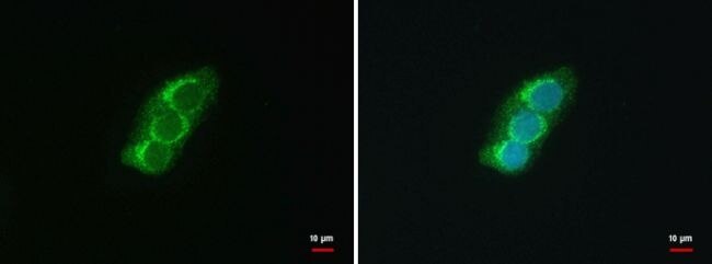
- Experimental details
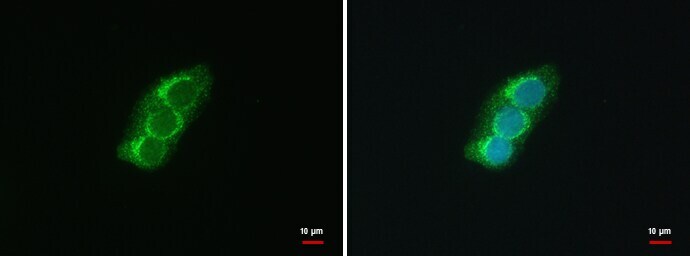
- SEC13 Polyclonal Antibody detects SEC13L1 protein at COP II vesicles by immunofluorescent analysis. Sample: HepG2 cells were fixed in 4% paraformaldehyde at RT for 15 min. Green: SEC13L1 protein stained by SEC13 Polyclonal Antibody (Product # PA5-21339) diluted at 1:500. Blue: Hoechst 33342 staining.
- Submitted by
- Invitrogen Antibodies (provider)
- Main image

- Experimental details
- SEC13 Polyclonal Antibody detects SEC13L1 protein at COP II vesicles by immunofluorescent analysis. Sample: HepG2 cells were fixed in 4% paraformaldehyde at RT for 15 min. Green: SEC13L1 protein stained by SEC13 Polyclonal Antibody (Product # PA5-21339) diluted at 1:500. Blue: Hoechst 33342 staining.

Supportive validation
- Submitted by
- Invitrogen Antibodies (provider)
- Main image

- Experimental details
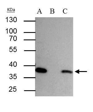
- SEC13L1 antibody immunoprecipitates SEC13L1 protein in IP experiments. IP Sample: HepG2 whole cell lysate/extract A : 30 µg whole cell lysate/extract of SEC13L1 protein expressing HepG2 cells B : Control with 2.5 µg of pre-immune rabbit IgG C : Immunoprecipitation of SEC13L1 by 2.5 µg of SEC13L1 antibody (Product # PA5-21339) 10% SDS-PAGE The immunoprecipitated SEC13L1 protein was detected by SEC13L1 antibody (Product # PA5-21339) diluted at 1:1,000. Anti-rabbit IgG (HRP) was used as a secondary reagent.
Supportive validation
- Submitted by
- Invitrogen Antibodies (provider)
- Main image
- Experimental details
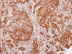
- Immunohistochemical analysis of paraffin-embedded A549 xenograft, using SEC13L1 (Product # PA5-21339) antibody at 1:100 dilution. Antigen Retrieval: Citrate buffer, pH 6.0, 15 min.

Supportive validation
- Submitted by
- Invitrogen Antibodies (provider)
- Main image

- Experimental details
- SEC13L1 antibody immunoprecipitates SEC13L1 protein in IP experiments. IP Sample: HepG2 whole cell lysate/extract A : 30 µg whole cell lysate/extract of SEC13L1 protein expressing HepG2 cells B : Control with 2.5 µg of pre-immune rabbit IgG C : Immunoprecipitation of SEC13L1 by 2.5 µg of SEC13L1 antibody (Product # PA5-21339) 10% SDS-PAGE The immunoprecipitated SEC13L1 protein was detected by SEC13L1 antibody (Product # PA5-21339) diluted at 1:1,000. Anti-rabbit IgG (HRP) was used as a secondary reagent.
