 Explore
 Validate
 Learn
Learn Western blot
 Immunocytochemistry
Antibody data
- Antibody Data
- Antigen structure
- References [24]
- Comments [0]
- Validations
- Immunocytochemistry [8]
- Immunohistochemistry [1]
- Other assay [10]
Submit
Validation data
Reference
Comment
Report error
- Product number
- MA1-720 - Provider product page

- Provider
- Invitrogen Antibodies
- Product name
- GRK1 Monoclonal Antibody (G8)
- Antibody type
- Monoclonal
- Antigen
- Other
- Description
- MA1-720 detects G-protein-associated rhodopsin kinase 1a (GRK1a) from human, mouse, rat, bovine and chicken tissues. MA1-720 has been successfully used in Western blot, immunofluorescence and immunoprecipitation procedures. By Western blot, this antibody detects an ~60 kDa protein representing GRK1a from bovine rod outer segments. Immunofluorescence staining of GRK1a in bovine retina with MA1-720 results in intense staining primarily in cone outer segments, but also in rod outer segments. Weak staining is observed in somata and synaptic terminals of cones and inner segments of rods. The MA1-720 immunogen is full length human GRK1. This antibody recognizes an epitope in the C-terminal region of GRK1a.
- Reactivity
- Human, Mouse, Rat, Bovine, Chicken/Avian
- Host
- Mouse
- Isotype
- IgG
- Antibody clone number
- G8
- Vial size
- 100 μg
- Concentration
- 1 mg/mL
- Storage
- -20°C, Avoid Freeze/Thaw Cycles
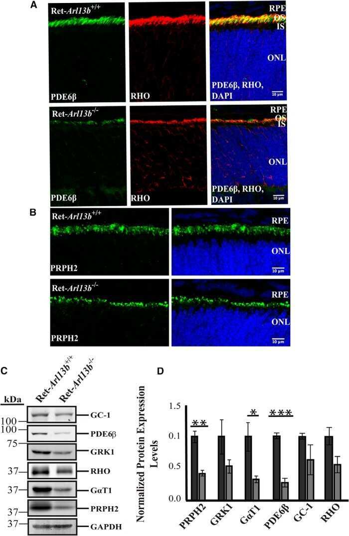
Submitted references ARL13B, a Joubert Syndrome-Associated Protein, Is Critical for Retinogenesis and Elaboration of Mouse Photoreceptor Outer Segments.
The age-regulating protein klotho is vital to sustain retinal function.
Modulation of mouse rod response decay by rhodopsin kinase and recoverin.
Tubby is required for trafficking G protein-coupled receptors to neuronal cilia.
Replacing the rod with the cone transducin subunit decreases sensitivity and accelerates response decay.
Control of rhodopsin's active lifetime by arrestin-1 expression in mammalian rods.
Background light produces a recoverin-dependent modulation of activated-rhodopsin lifetime in mouse rods.
Arrestin competition influences the kinetics and variability of the single-photon responses of mammalian rod photoreceptors.
CREB1/ATF1 activation in photoreceptor degeneration and protection.
High levels of retinal membrane docosahexaenoic acid increase susceptibility to stress-induced degeneration.
Overexpression of rhodopsin alters the structure and photoresponse of rod photoreceptors.
Flupirtine attenuates sodium nitroprusside-induced damage to retinal photoreceptors, in situ.
Differentiation of embryonic stem cells to retinal cells in vitro.
The beta-adrenergic receptor antagonist metipranolol blunts zinc-induced photoreceptor and RPE apoptosis.
RGS expression rate-limits recovery of rod photoresponses.
Functional characterization of mouse RDH11 as a retinol dehydrogenase involved in dark adaptation in vivo.
Evaluation of the 17-kDa prenyl-binding protein as a regulatory protein for phototransduction in retinal photoreceptors.
Transducin activation state controls its light-dependent translocation in rod photoreceptors.
Leber congenital amaurosis linked to AIPL1: a mouse model reveals destabilization of cGMP phosphodiesterase.
The bovine iris-ciliary epithelium expresses components of rod phototransduction.
Cone deactivation kinetics and GRK1/GRK7 expression in enhanced S cone syndrome caused by mutations in NR2E3.
A novel form of rhodopsin kinase from chicken retina and pineal gland.
Phosphorylation of photolyzed rhodopsin is calcium-insensitive in retina permeabilized by alpha-toxin.
Phosphorylation of photolyzed rhodopsin is calcium-insensitive in retina permeabilized by alpha-toxin.
Dilan TL, Moye AR, Salido EM, Saravanan T, Kolandaivelu S, Goldberg AFX, Ramamurthy V
The Journal of neuroscience : the official journal of the Society for Neuroscience 2019 Feb 20;39(8):1347-1364
The Journal of neuroscience : the official journal of the Society for Neuroscience 2019 Feb 20;39(8):1347-1364
The age-regulating protein klotho is vital to sustain retinal function.
Reish NJ, Maltare A, McKeown AS, Laszczyk AM, Kraft TW, Gross AK, King GD
Investigative ophthalmology & visual science 2013 Oct 11;54(10):6675-85
Investigative ophthalmology & visual science 2013 Oct 11;54(10):6675-85
Modulation of mouse rod response decay by rhodopsin kinase and recoverin.
Chen CK, Woodruff ML, Chen FS, Chen Y, Cilluffo MC, Tranchina D, Fain GL
The Journal of neuroscience : the official journal of the Society for Neuroscience 2012 Nov 7;32(45):15998-6006
The Journal of neuroscience : the official journal of the Society for Neuroscience 2012 Nov 7;32(45):15998-6006
Tubby is required for trafficking G protein-coupled receptors to neuronal cilia.
Sun X, Haley J, Bulgakov OV, Cai X, McGinnis J, Li T
Cilia 2012 Nov 1;1(1):21
Cilia 2012 Nov 1;1(1):21
Replacing the rod with the cone transducin subunit decreases sensitivity and accelerates response decay.
Chen CK, Woodruff ML, Chen FS, Shim H, Cilluffo MC, Fain GL
The Journal of physiology 2010 Sep 1;588(Pt 17):3231-41
The Journal of physiology 2010 Sep 1;588(Pt 17):3231-41
Control of rhodopsin's active lifetime by arrestin-1 expression in mammalian rods.
Gross OP, Burns ME
The Journal of neuroscience : the official journal of the Society for Neuroscience 2010 Mar 3;30(9):3450-7
The Journal of neuroscience : the official journal of the Society for Neuroscience 2010 Mar 3;30(9):3450-7
Background light produces a recoverin-dependent modulation of activated-rhodopsin lifetime in mouse rods.
Chen CK, Woodruff ML, Chen FS, Chen D, Fain GL
The Journal of neuroscience : the official journal of the Society for Neuroscience 2010 Jan 27;30(4):1213-20
The Journal of neuroscience : the official journal of the Society for Neuroscience 2010 Jan 27;30(4):1213-20
Arrestin competition influences the kinetics and variability of the single-photon responses of mammalian rod photoreceptors.
Doan T, Azevedo AW, Hurley JB, Rieke F
The Journal of neuroscience : the official journal of the Society for Neuroscience 2009 Sep 23;29(38):11867-79
The Journal of neuroscience : the official journal of the Society for Neuroscience 2009 Sep 23;29(38):11867-79
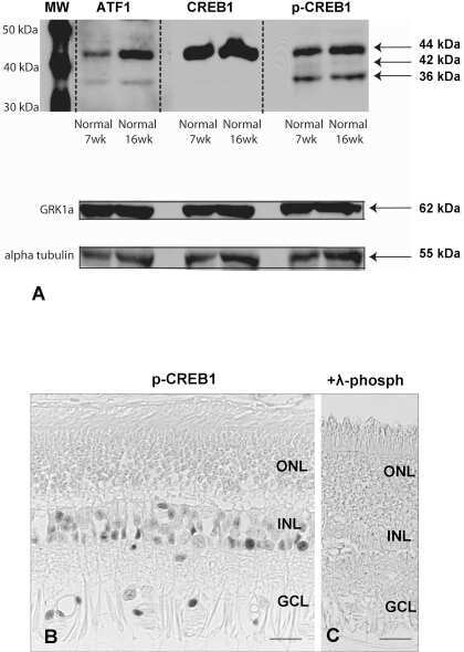
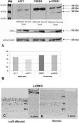
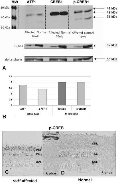
CREB1/ATF1 activation in photoreceptor degeneration and protection.
Beltran WA, Allore HG, Johnson E, Towle V, Tao W, Acland GM, Aguirre GD, Zeiss CJ
Investigative ophthalmology & visual science 2009 Nov;50(11):5355-63
Investigative ophthalmology & visual science 2009 Nov;50(11):5355-63
High levels of retinal membrane docosahexaenoic acid increase susceptibility to stress-induced degeneration.
Tanito M, Brush RS, Elliott MH, Wicker LD, Henry KR, Anderson RE
Journal of lipid research 2009 May;50(5):807-19
Journal of lipid research 2009 May;50(5):807-19
Overexpression of rhodopsin alters the structure and photoresponse of rod photoreceptors.
Wen XH, Shen L, Brush RS, Michaud N, Al-Ubaidi MR, Gurevich VV, Hamm HE, Lem J, Dibenedetto E, Anderson RE, Makino CL
Biophysical journal 2009 Feb;96(3):939-50
Biophysical journal 2009 Feb;96(3):939-50
Flupirtine attenuates sodium nitroprusside-induced damage to retinal photoreceptors, in situ.
Fawcett RJ, Osborne NN
Brain research bulletin 2007 Jul 12;73(4-6):278-88
Brain research bulletin 2007 Jul 12;73(4-6):278-88
Differentiation of embryonic stem cells to retinal cells in vitro.
Zhao X, Liu J, Ahmad I
Methods in molecular biology (Clifton, N.J.) 2006;330:401-16
Methods in molecular biology (Clifton, N.J.) 2006;330:401-16
The beta-adrenergic receptor antagonist metipranolol blunts zinc-induced photoreceptor and RPE apoptosis.
Osborne NN, Wood JP
Investigative ophthalmology & visual science 2006 Jul;47(7):3178-86
Investigative ophthalmology & visual science 2006 Jul;47(7):3178-86
RGS expression rate-limits recovery of rod photoresponses.
Krispel CM, Chen D, Melling N, Chen YJ, Martemyanov KA, Quillinan N, Arshavsky VY, Wensel TG, Chen CK, Burns ME
Neuron 2006 Aug 17;51(4):409-16
Neuron 2006 Aug 17;51(4):409-16
Functional characterization of mouse RDH11 as a retinol dehydrogenase involved in dark adaptation in vivo.
Kasus-Jacobi A, Ou J, Birch DG, Locke KG, Shelton JM, Richardson JA, Murphy AJ, Valenzuela DM, Yancopoulos GD, Edwards AO
The Journal of biological chemistry 2005 May 27;280(21):20413-20
The Journal of biological chemistry 2005 May 27;280(21):20413-20
Evaluation of the 17-kDa prenyl-binding protein as a regulatory protein for phototransduction in retinal photoreceptors.
Norton AW, Hosier S, Terew JM, Li N, Dhingra A, Vardi N, Baehr W, Cote RH
The Journal of biological chemistry 2005 Jan 14;280(2):1248-56
The Journal of biological chemistry 2005 Jan 14;280(2):1248-56
Transducin activation state controls its light-dependent translocation in rod photoreceptors.
Kerov V, Chen D, Moussaif M, Chen YJ, Chen CK, Artemyev NO
The Journal of biological chemistry 2005 Dec 9;280(49):41069-76
The Journal of biological chemistry 2005 Dec 9;280(49):41069-76
Leber congenital amaurosis linked to AIPL1: a mouse model reveals destabilization of cGMP phosphodiesterase.
Ramamurthy V, Niemi GA, Reh TA, Hurley JB
Proceedings of the National Academy of Sciences of the United States of America 2004 Sep 21;101(38):13897-902
Proceedings of the National Academy of Sciences of the United States of America 2004 Sep 21;101(38):13897-902
The bovine iris-ciliary epithelium expresses components of rod phototransduction.
Ghosh S, Salvador-Silva M, Coca-Prados M
Neuroscience letters 2004 Nov 3;370(1):7-12
Neuroscience letters 2004 Nov 3;370(1):7-12
Cone deactivation kinetics and GRK1/GRK7 expression in enhanced S cone syndrome caused by mutations in NR2E3.
Cideciyan AV, Jacobson SG, Gupta N, Osawa S, Locke KG, Weiss ER, Wright AF, Birch DG, Milam AH
Investigative ophthalmology & visual science 2003 Mar;44(3):1268-74
Investigative ophthalmology & visual science 2003 Mar;44(3):1268-74
A novel form of rhodopsin kinase from chicken retina and pineal gland.
Zhao X, Yokoyama K, Whitten ME, Huang J, Gelb MH, Palczewski K
FEBS letters 1999 Jul 2;454(1-2):115-21
FEBS letters 1999 Jul 2;454(1-2):115-21
Phosphorylation of photolyzed rhodopsin is calcium-insensitive in retina permeabilized by alpha-toxin.
Otto-Bruc AE, Fariss RN, Van Hooser JP, Palczewski K
Proceedings of the National Academy of Sciences of the United States of America 1998 Dec 8;95(25):15014-9
Proceedings of the National Academy of Sciences of the United States of America 1998 Dec 8;95(25):15014-9
No comments: Submit comment
Supportive validation
- Submitted by
- Invitrogen Antibodies (provider)
- Main image

- Experimental details
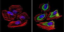
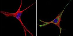
- Immunofluorescent analysis of Rhodopsin Kinase 1a (green) in human cells. Formalin-fixed cells were permeabilized with 0.1% Triton X-100 in TBS for 5-10 minutes at room temperature and blocked with 3% BSA-PBS for 30 minutes at room temperature. Cells were probed with a Rhodopsin Kinase 1a Monclonal Antibody (G8) (Product # MA1-720) at a dilution of 1:100 and incubated overnight in a humidified chamber. Cells were washed with PBST and incubated with a DyLight-conjugated secondary antibody for 45 minutes at room temperature in the dark. F-actin (red) was stained with a fluorescent phalloidin and nuclei (blue) were stained with DAPI. Images were taken at a 60X magnification.
- Submitted by
- Invitrogen Antibodies (provider)
- Main image
- Experimental details
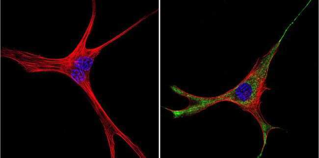
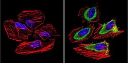
- Immunofluorescent analysis of Rhodopsin Kinase 1a (green) in Hela cells. Formalin-fixed cells were permeabilized with 0.1% Triton X-100 in TBS for 5-10 minutes at room temperature and blocked with 3% BSA-PBS for 30 minutes at room temperature. Cells were probed with a Rhodopsin Kinase 1a Monclonal Antibody (G8) (Product # MA1-720) at a dilution of 1:200 and incubated overnight in a humidified chamber. Cells were washed with PBST and incubated with a DyLight-conjugated secondary antibody for 45 minutes at room temperature in the dark. F-actin (red) was stained with a fluorescent phalloidin and nuclei (blue) were stained with DAPI. Images were taken at a 60X magnification.

- Submitted by
- Invitrogen Antibodies (provider)
- Main image
- Experimental details
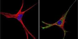
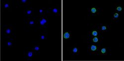
- Immunofluorescent analysis of Rhodopsin Kinase 1a (green) in murine cells. Formalin-fixed cells were permeabilized with 0.1% Triton X-100 in TBS for 5-10 minutes at room temperature and blocked with 3% BSA-PBS for 30 minutes at room temperature. Cells were probed with a Rhodopsin Kinase 1a Monclonal Antibody (G8) (Product # MA1-720) at a dilution of 1:200 and incubated overnight in a humidified chamber. Cells were washed with PBST and incubated with a DyLight-conjugated secondary antibody for 45 minutes at room temperature in the dark. F-actin (red) was stained with a fluorescent phalloidin and nuclei (blue) were stained with DAPI. Images were taken at a 60X magnification.
- Submitted by
- Invitrogen Antibodies (provider)
- Main image
- Experimental details
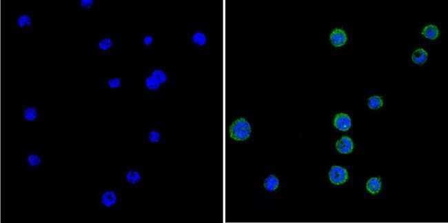
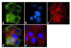
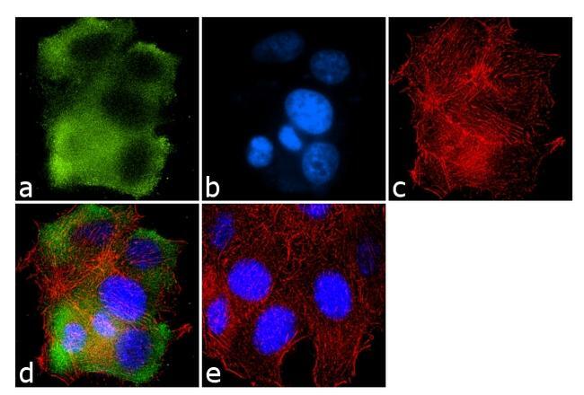
- Immunofluorescence analysis of Rhodopsin Kinase 1a was done on 70% confluent log phase MCF-7 cells. The cells were fixed with 4% paraformaldehyde for 10 minutes, permeabilized with 0.1% Triton™ X-100 for 10 minutes, and blocked with 1% BSA for 1 hour at room temperature. The cells were labeled with Rhodopsin Kinase 1a (G8) Mouse Monoclonal Antibody (Product # MA1-720) at 2 µg/mL in 0.1% BSA and incubated for 3 hours at room temperature and then labeled with Goat anti-Mouse IgG (H+L) Superclonal™ Secondary Antibody, Alexa Fluor® 488 conjugate (Product # A28175) at a dilution of 1:2000 for 45 minutes at room temperature (Panel a: green). Nuclei (Panel b: blue) were stained with SlowFade® Gold Antifade Mountant with DAPI (Product # S36938). F-actin (Panel c: red) was stained with Alexa Fluor® 555 Rhodamine Phalloidin (Product # R415, 1:300). Panel d is a merged image showing Cytoplasmic localization. Panel e is a no primary antibody control. The images were captured at 60X magnification.

- Submitted by
- Invitrogen Antibodies (provider)
- Main image
- Experimental details
- Immunofluorescent analysis of Rhodopsin Kinase 1a (green) in human cells. Formalin-fixed cells were permeabilized with 0.1% Triton X-100 in TBS for 5-10 minutes at room temperature and blocked with 3% BSA-PBS for 30 minutes at room temperature. Cells were probed with a Rhodopsin Kinase 1a Monclonal Antibody (G8) (Product # MA1-720) at a dilution of 1:100 and incubated overnight in a humidified chamber. Cells were washed with PBST and incubated with a DyLight-conjugated secondary antibody for 45 minutes at room temperature in the dark. F-actin (red) was stained with a fluorescent phalloidin and nuclei (blue) were stained with DAPI. Images were taken at a 60X magnification.

- Submitted by
- Invitrogen Antibodies (provider)
- Main image
- Experimental details
- Immunofluorescent analysis of Rhodopsin Kinase 1a (green) in Hela cells. Formalin-fixed cells were permeabilized with 0.1% Triton X-100 in TBS for 5-10 minutes at room temperature and blocked with 3% BSA-PBS for 30 minutes at room temperature. Cells were probed with a Rhodopsin Kinase 1a Monclonal Antibody (G8) (Product # MA1-720) at a dilution of 1:200 and incubated overnight in a humidified chamber. Cells were washed with PBST and incubated with a DyLight-conjugated secondary antibody for 45 minutes at room temperature in the dark. F-actin (red) was stained with a fluorescent phalloidin and nuclei (blue) were stained with DAPI. Images were taken at a 60X magnification.

- Submitted by
- Invitrogen Antibodies (provider)
- Main image
- Experimental details
- Immunofluorescent analysis of Rhodopsin Kinase 1a (green) in murine cells. Formalin-fixed cells were permeabilized with 0.1% Triton X-100 in TBS for 5-10 minutes at room temperature and blocked with 3% BSA-PBS for 30 minutes at room temperature. Cells were probed with a Rhodopsin Kinase 1a Monclonal Antibody (G8) (Product # MA1-720) at a dilution of 1:200 and incubated overnight in a humidified chamber. Cells were washed with PBST and incubated with a DyLight-conjugated secondary antibody for 45 minutes at room temperature in the dark. F-actin (red) was stained with a fluorescent phalloidin and nuclei (blue) were stained with DAPI. Images were taken at a 60X magnification.

- Submitted by
- Invitrogen Antibodies (provider)
- Main image

- Experimental details
- Immunofluorescence analysis of Rhodopsin Kinase 1a was done on 70% confluent log phase MCF-7 cells. The cells were fixed with 4% paraformaldehyde for 10 minutes, permeabilized with 0.1% Triton™ X-100 for 10 minutes, and blocked with 1% BSA for 1 hour at room temperature. The cells were labeled with Rhodopsin Kinase 1a (G8) Mouse Monoclonal Antibody (Product # MA1-720) at 2 µg/mL in 0.1% BSA and incubated for 3 hours at room temperature and then labeled with Goat anti-Mouse IgG (H+L) Superclonal™ Secondary Antibody, Alexa Fluor® 488 conjugate (Product # A28175) at a dilution of 1:2000 for 45 minutes at room temperature (Panel a: green). Nuclei (Panel b: blue) were stained with SlowFade® Gold Antifade Mountant with DAPI (Product # S36938). F-actin (Panel c: red) was stained with Alexa Fluor® 555 Rhodamine Phalloidin (Product # R415, 1:300). Panel d is a merged image showing Cytoplasmic localization. Panel e is a no primary antibody control. The images were captured at 60X magnification.
Supportive validation
- Submitted by
- Invitrogen Antibodies (provider)
- Main image
- Experimental details
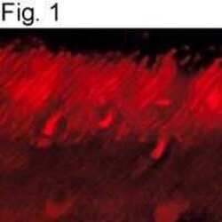
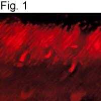
- Immunolocalization of GRK1a in bovine retina using Product # MA1-720.
Supportive validation
- Submitted by
- Invitrogen Antibodies (provider)
- Main image
- Experimental details
- NULL
- Submitted by
- Invitrogen Antibodies (provider)
- Main image
- Experimental details
- NULL
- Submitted by
- Invitrogen Antibodies (provider)
- Main image
- Experimental details
- NULL

- Submitted by
- Invitrogen Antibodies (provider)
- Main image

- Experimental details
- NULL
- Submitted by
- Invitrogen Antibodies (provider)
- Main image
- Experimental details
- NULL
- Submitted by
- Invitrogen Antibodies (provider)
- Main image
- Experimental details
- NULL
- Submitted by
- Invitrogen Antibodies (provider)
- Main image
- Experimental details
- NULL
- Submitted by
- Invitrogen Antibodies (provider)
- Main image
- Experimental details
- NULL

- Submitted by
- Invitrogen Antibodies (provider)
- Main image
- Experimental details
- NULL
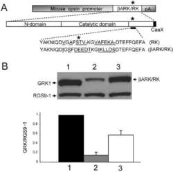
- Submitted by
- Invitrogen Antibodies (provider)
- Main image

- Experimental details
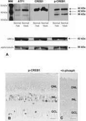
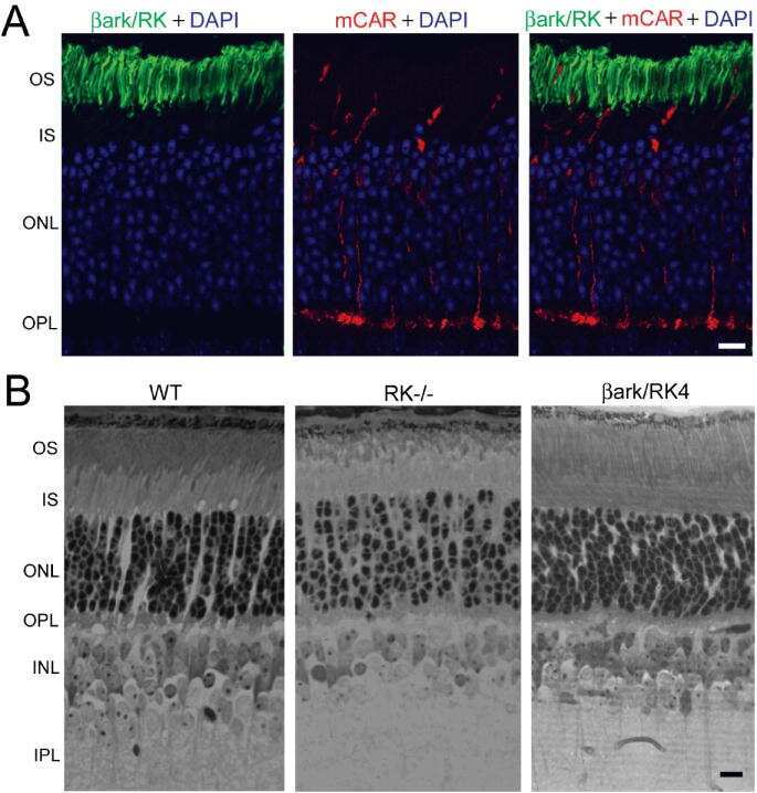
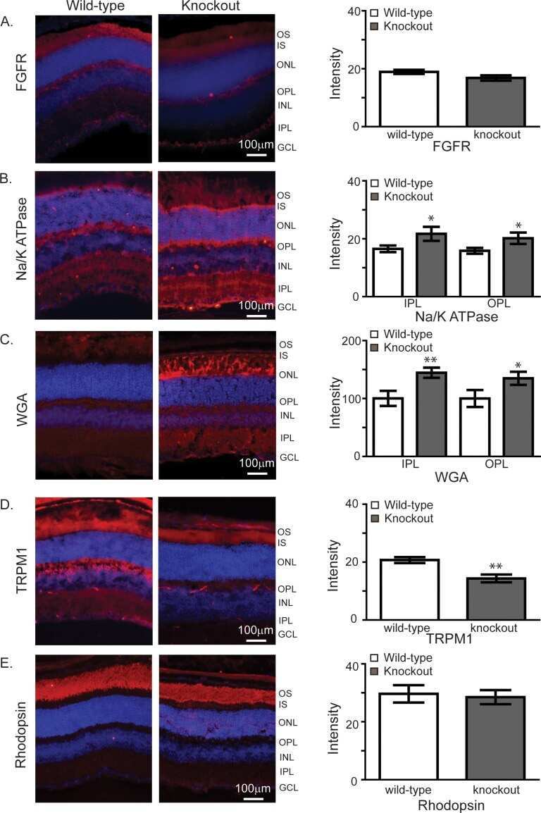
- Figure 1 Immunofluorescence analyses of the tubby mutant retina. (A) The WT tubby protein migrates at approximately 60 kDa by immunoblotting, but tubby protein expression is absent in the mutant tissues indicating that the mutation is a functional allele. The lower panel shows the actin immunoblotting results as a loading control. (B) Rhodopsin is localized primarily in photoreceptor outer segments in WT but is severely mislocalized to the cell body in the tubby mutant at one month of age. Similarly, blue and green cone opsins are normally localized to the cone outer segments in WT but are also mislocalized to the cell body and synaptic terminals in the tubby mutant retina. (C) In contrast to rhodopsin and cone opsins, peripherin/RDS and GRK, which are also integral membrane proteins, do not show mislocalization in the tubby mutant. Rod PDE and RP1 protein, which are membrane and cytoskeleton associated proteins, respectively, also manifest normal outer segment localization. IS, inner segment; ONL, outer (photoreceptor) nuclear layer; OPL, outer plexiform layer; OS, outer segment. Cell nuclei were counter stained blue by DAPI.
