Explore
 Validate
 Learn
Learn Western blot
 ELISA
ELISA Immunocytochemistry
Antibody data
- Antibody Data
- Antigen structure
- References [6]
- Comments [0]
- Validations
- Immunocytochemistry [2]
- Immunohistochemistry [1]
- Flow cytometry [1]
- Other assay [2]
Submit
Validation data
Reference
Comment
Report error
- Product number
- 36-6200 - Provider product page

- Provider
- Invitrogen Antibodies
- Product name
- Syndecan 2 Polyclonal Antibody
- Antibody type
- Polyclonal
- Antigen
- Synthetic peptide
- Reactivity
- Human
- Host
- Rabbit
- Isotype
- IgG
- Vial size
- 100 μg
- Concentration
- 0.25 mg/mL
- Storage
- -20°C
Submitted references Plasma membrane proteoglycans syndecan-2 and syndecan-4 engage with EGFR and RON kinase to sustain carcinoma cell cycle progression.
Heparan sulfate proteoglycans (HSPGs) and chondroitin sulfate proteoglycans (CSPGs) function as endocytic receptors for an internalizing anti-nucleic acid antibody.
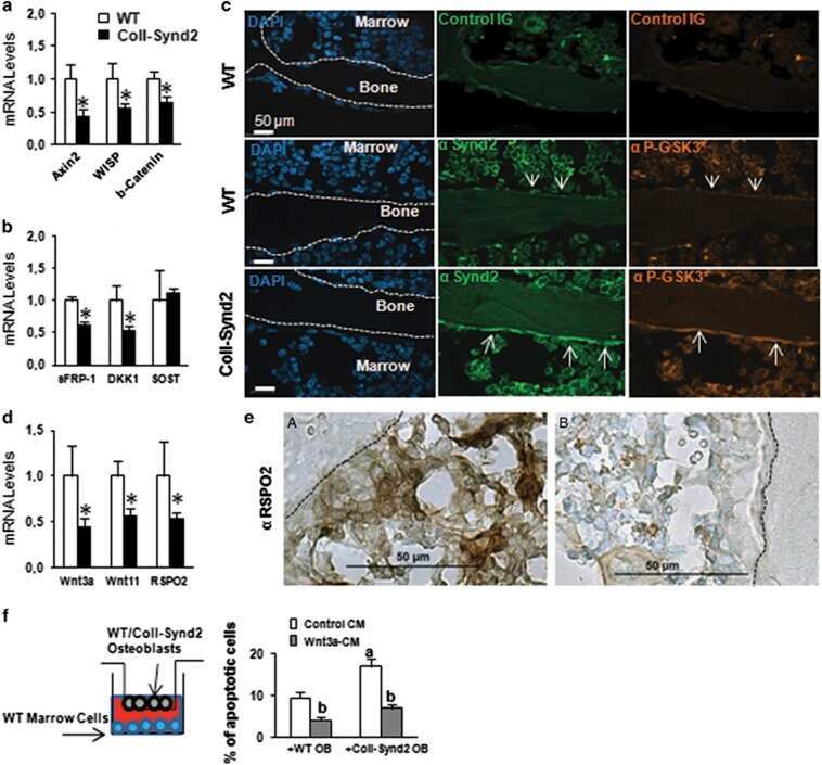
Osteoblastic heparan sulfate glycosaminoglycans control bone remodeling by regulating Wnt signaling and the crosstalk between bone surface and marrow cells.
Syndecan-2 is a key regulator of transforming growth factor beta 2/Smad2-mediated adhesion in fibrosarcoma cells.
Differences in heparan sulfate production in cervical fibroblast cultures from women undergoing term and preterm delivery.
CCN2 (connective tissue growth factor) promotes fibroblast adhesion to fibronectin.
Beauvais DM, Nelson SE, Adams KM, Stueven NA, Jung O, Rapraeger AC
The Journal of biological chemistry 2022 Jun;298(6):102029
The Journal of biological chemistry 2022 Jun;298(6):102029
Heparan sulfate proteoglycans (HSPGs) and chondroitin sulfate proteoglycans (CSPGs) function as endocytic receptors for an internalizing anti-nucleic acid antibody.
Park H, Kim M, Kim HJ, Lee Y, Seo Y, Pham CD, Lee J, Byun SJ, Kwon MH
Scientific reports 2017 Oct 30;7(1):14373
Scientific reports 2017 Oct 30;7(1):14373
Osteoblastic heparan sulfate glycosaminoglycans control bone remodeling by regulating Wnt signaling and the crosstalk between bone surface and marrow cells.
Mansouri R, Jouan Y, Hay E, Blin-Wakkach C, Frain M, Ostertag A, Le Henaff C, Marty C, Geoffroy V, Marie PJ, Cohen-Solal M, Modrowski D
Cell death & disease 2017 Jun 29;8(6):e2902
Cell death & disease 2017 Jun 29;8(6):e2902
Syndecan-2 is a key regulator of transforming growth factor beta 2/Smad2-mediated adhesion in fibrosarcoma cells.
Mytilinaiou M, Bano A, Nikitovic D, Berdiaki A, Voudouri K, Kalogeraki A, Karamanos NK, Tzanakakis GN
IUBMB life 2013 Feb;65(2):134-43
IUBMB life 2013 Feb;65(2):134-43
Differences in heparan sulfate production in cervical fibroblast cultures from women undergoing term and preterm delivery.
Akerud A, Dubicke A, Sennstrom M, Ekman-Ordeberg G, Malmstrom A
Acta obstetricia et gynecologica Scandinavica 2008;87(11):1220-8
Acta obstetricia et gynecologica Scandinavica 2008;87(11):1220-8
CCN2 (connective tissue growth factor) promotes fibroblast adhesion to fibronectin.
Chen Y, Abraham DJ, Shi-Wen X, Pearson JD, Black CM, Lyons KM, Leask A
Molecular biology of the cell 2004 Dec;15(12):5635-46
Molecular biology of the cell 2004 Dec;15(12):5635-46
No comments: Submit comment
Supportive validation
- Submitted by
- Invitrogen Antibodies (provider)
- Main image

- Experimental details
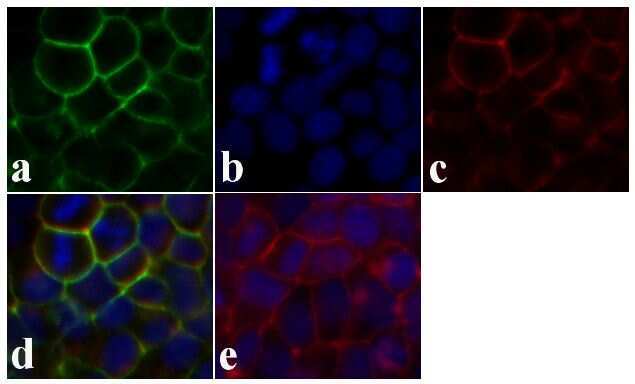
- Immunofluorescence analysis of Syndecan-2 was done on 90% confluent log phase Caco-2 cells. The cells were fixed with 4% paraformaldehyde for 15 minutes, permeabilized with 0.25% Triton X-100 for 10 minutes, and blocked with 5% BSA for 1 hour at room temperature. The cells were labeled with Syndecan-2 Rabbit polyclonal Antibody (Product # 36-6200) at 2 µg/mL in 1% BSA and incubated for 3 hours at room temperature and then labeled with Alexa Fluor 488 Goat Anti-Rabbit IgG Secondary Antibody (Product # A-11008) at a dilution of 1:400 for 30 minutes at room temperature (Panel a: green). Nuclei (Panel b: blue) were stained with SlowFade® Gold Antifade Mountant DAPI (Product # S36938). F-actin (Panel c: red) was stained with Alexa Fluor 594 Phalloidin (Product # A12381). Panel d is a merged image showing junctional localization. Panel e shows no primary antibody control. The images were captured at 20X magnification.
- Submitted by
- Invitrogen Antibodies (provider)
- Main image
- Experimental details
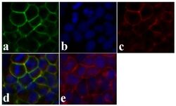
- Immunofluorescence analysis of Syndecan-2 was done on 90% confluent log phase Caco-2 cells. The cells were fixed with 4% paraformaldehyde for 15 minutes, permeabilized with 0.25% Triton X-100 for 10 minutes, and blocked with 5% BSA for 1 hour at room temperature. The cells were labeled with Syndecan-2 Rabbit polyclonal Antibody (Product # 36-6200) at 2 µg/mL in 1% BSA and incubated for 3 hours at room temperature and then labeled with Alexa Fluor 488 Goat Anti-Rabbit IgG Secondary Antibody (Product # A-11008) at a dilution of 1:400 for 30 minutes at room temperature (Panel a: green). Nuclei (Panel b: blue) were stained with SlowFade® Gold Antifade Mountant DAPI (Product # S36938). F-actin (Panel c: red) was stained with Alexa Fluor 594 Phalloidin (Product # A12381). Panel d is a merged image showing junctional localization. Panel e shows no primary antibody control. The images were captured at 20X magnification.

Supportive validation
- Submitted by
- Invitrogen Antibodies (provider)
- Main image
- Experimental details
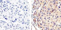
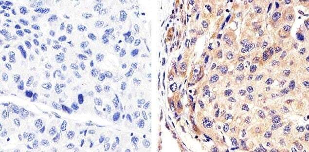
- Immunohistochemistry analysis ofSyndecan-2 showing staining in the cytoplasm of paraffin-embedded human hepatocellular carcinoma tissue (right) compared to a negative control without primary antibody (left). To expose target proteins, antigen retrieval was performed using 10mM sodium citrate (pH 6.0), microwaved for 8-15 min. Following antigen retrieval, tissues were blocked in 3% H2O2-methanol for 15 min at room temperature, washed with ddH2O and PBS, and then probed with a Syndecan-2 polyclonal antibody (Product # 36-6200) diluted in 3% BSA-PBS at a dilution of 1:20 overnight at 4°C in a humidified chamber. Tissues were washed extensively in PBST and detection was performed using an HRP-conjugated secondary antibody followed by colorimetric detection using a DAB kit. Tissues were counterstained with hematoxylin and dehydrated with ethanol and xylene to prep for mounting.
Supportive validation
- Submitted by
- Invitrogen Antibodies (provider)
- Main image
- Experimental details
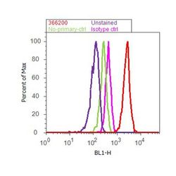
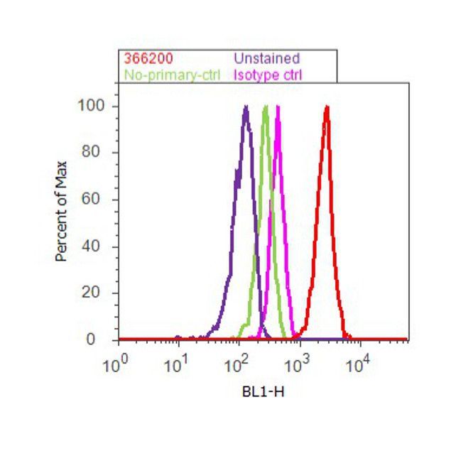
- Flow cytometry analysis of Syndecan-2 was done on HeLa cells. Cells were fixed with 70% ethanol for 10 minutes, permeabilized with 0.25% Tritonª X-100 for 20 minutes, and blocked with 5% BSA for 30 minutes at room temperature. Cells were labeled with Syndecan-2 Rabbit Polyclonal Antibody (366200, red histogram) or with rabbit isotype control (pink histogram) at 3-5 µg/million cells in 2.5% BSA. After incubation at room temperature for 2 hours, the cells were labeled with Alexa Fluor¨ 488 Goat Anti-Rabbit Secondary Antibody (A11008) at a dilution of 1:400 for 30 minutes at room temperature. The representative 10,000 cells were acquired and analyzed for each sample using an Attune¨ Acoustic Focusing Cytometer. The purple histogram represents unstained control cells and the green histogram represents no-primary-antibody control.
Supportive validation
- Submitted by
- Invitrogen Antibodies (provider)
- Main image
- Experimental details
- NULL
- Submitted by
- Invitrogen Antibodies (provider)
- Main image
- Experimental details
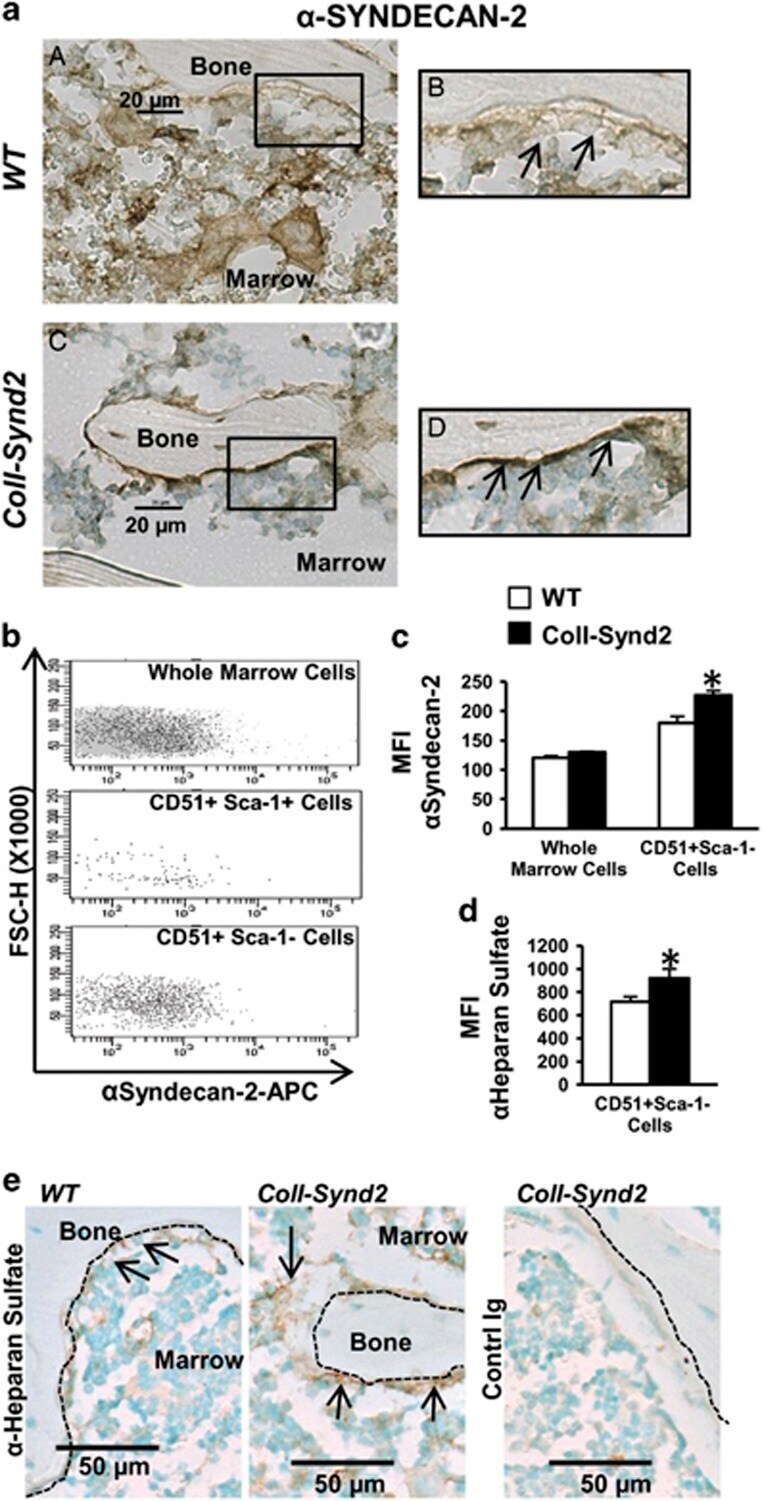
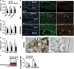
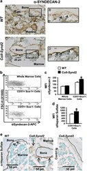
- Figure 1 Syndecan-2 overexpression enriched bone surface with heparan sulfate glycosaminoglycans. ( a ) Immunohistological analysis of syndecan-2 in paraffin-embedded vertebra from 2-month-old WT ( a and b ) or ColI-Synd2 ( c and d ) mice. ( b ) Flow cytometry analysis of syndecan-2 expression. Marrow cells from 6-week-old mice were flushed from long bones and labeled with surface markers to select osteoblast precursors (CD51 + Sca-1 + cells) or osteoblasts (CD51 + Sca-1 - cells). Fluorescence intensity versus structural parameter of the cells (FSC-H) was plotted. ( c ) The median fluorescence intensity (MFI) of syndecan-2 labeling was recorded in different cell populations from WT or ColI-Synd2 mice. Data are mean+-S.E.M. ( n =7 WT and three ColI-Synd2 mice). ( d ) The MFI of heparan sulfate labeling was recorded in CD51 + Sca-1 - cells from the marrow of WT or ColI-Synd2 mice. Data are mean+-S.E.M. ( n =4 WT and four ColI-Synd2 mice). ( e ) Immunohistological analysis of heparan sulfate chains in paraffin-embedded vertebra from 2-month-old WT or ColI-Synd2 mice. The photos presented are representative views of syndecan-2 or heparan sulfate staining. Arrows indicate syndecan-2- or heparan sulfate-expressing cells. Dashed lines indicate bone surfaces. *Indicates significant difference between WT and ColI-Synd2 ( P