 Explore
 Validate
 Learn
Learn Western blot
Antibody data
- Antibody Data
- Antigen structure
- References [1]
- Comments [0]
- Validations
- Western blot [1]
- Immunohistochemistry [1]
- Flow cytometry [1]
Submit
Validation data
Reference
Comment
Report error
- Product number
- AF6585 - Provider product page

- Provider
- R&D Systems
- Product name
- Mouse Syndecan-2/CD362 Antibody
- Antibody type
- Polyclonal
- Description
- Antigen Affinity-purified. Detects mouse Syndecan-2/CD362 in direct ELISAs and Western blots. In direct ELISAs, less than 1% cross-reactivity with recombinant human Syndecan-2/CD362 is observed.
- Reactivity
- Mouse
- Host
- Sheep
- Conjugate
- Unconjugated
- Antigen sequence
P43407- Isotype
- IgG
- Vial size
- 100 ug
- Concentration
- LYOPH
- Storage
- Use a manual defrost freezer and avoid repeated freeze-thaw cycles. 12 months from date of receipt, -20 to -70 °C as supplied. 1 month, 2 to 8 °C under sterile conditions after reconstitution. 6 months, -20 to -70 °C under sterile conditions after reconstitution.
Submitted references RKIP and HMGA2 regulate breast tumor survival and metastasis through lysyl oxidase and syndecan-2.
Sun M, Gomes S, Chen P, Frankenberger CA, Sankarasharma D, Chung CH, Chada KK, Rosner MR
Oncogene 2014 Jul 3;33(27):3528-37
Oncogene 2014 Jul 3;33(27):3528-37
No comments: Submit comment
Supportive validation
- Submitted by
- R&D Systems (provider)
- Main image

- Experimental details
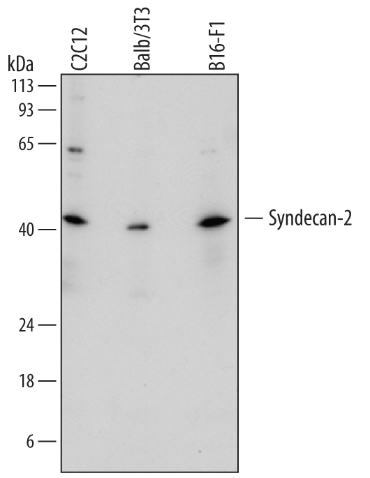
- Detection of Mouse Syndecan-2/CD362 by Western Blot. Western blot shows lysates of C2C12 mouse myoblast cell line, Balb/3T3 mouse embryonic fibroblast cell line, and B16-F1 mouse melanoma cell line. PVDF membrane was probed with 1 µg/mL of Sheep Anti-Mouse Syndecan-2/CD362 Antigen Affinity-purified Polyclonal Antibody (Catalog # AF6585) followed by HRP-conjugated Anti-Sheep IgG Secondary Antibody (Catalog # HAF016). A specific band was detected for Syndecan-2/CD362 at approximately 42 kDa (as indicated). This experiment was conducted under reducing conditions and using Immunoblot Buffer Group 1.
Supportive validation
- Submitted by
- R&D Systems (provider)
- Main image

- Experimental details
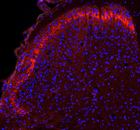
- Syndecan-2/CD362 in Mouse Spinal Cord. Syndecan-2/CD362 was detected in perfusion fixed frozen sections of mouse spinal cord using Sheep Anti-Mouse Syndecan-2/CD362 Antigen Affinity-purified Polyclonal Antibody (Catalog # AF6585) at 1.7 µg/mL overnight at 4 °C. Tissue was stained using the NorthernLights™ 557-conjugated Anti-Sheep IgG Secondary Antibody (red; Catalog # NL010) and counterstained with DAPI (blue). Specific staining was localized to the dorsal horn. View our protocol for Fluorescent IHC Staining of Frozen Tissue Sections.
Supportive validation
- Submitted by
- R&D Systems (provider)
- Main image
- Experimental details
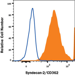
- Detection of Syndecan-2/CD362 in NIH-3T3 Mouse Cell Line by Flow Cytometry. NIH-3T3 mouse cell line was stained with Sheep Anti-Mouse Syndecan-2/CD362 Antigen Affinity-purified Polyclonal Antibody (Catalog # AF6585, filled histogram) or control antibody (Catalog # 5-001-A, open histogram), followed by Allophycocyanin-conjugated Anti-Sheep IgG Secondary Antibody (Catalog # F0127). View our protocol for Staining Membrane-associated Proteins.
