Explore
 Validate
 Learn
Learn Western blot
 ELISA
ELISAAntibody data
- Antibody Data
- Antigen structure
- References [1]
- Comments [0]
- Validations
- Western blot [1]
Submit
Validation data
Reference
Comment
Report error
- Product number
- A01601 - Provider product page

- Provider
- Boster Biological Technology
- Product name
- Anti-CYP7A1 Antibody Picoband™
- Antibody type
- Polyclonal
- Description
- Rabbit IgG polyclonal antibody for CYP7A1 detection. Tested with WB, FCM, Direct ELISA in Human;Mouse;Rat;Monkey.
- Reactivity
- Human, Mouse, Rat, Simian
- Host
- Rabbit
- Vial size
- 100μg/vial
- Concentration
- Add 0.2ml of distilled water will yield a concentration of 500ug/ml.
- Storage
- At -20°C for one year. After reconstitution, at 4°C for one month. It can also be aliquoted and stored frozen at -20°C for a longer time. Avoid repeated freezing and thawing.
- Handling
- Add 0.2ml of distilled water will yield a concentration of 500ug/ml.
Submitted references Cellular Accumulation and Toxic Effects of Bile Acids in Cyclosporine A-Treated HepaRG Hepatocytes.
Sharanek A, Burban A, Humbert L, Bachour-El Azzi P, Felix-Gomes N, Rainteau D, Guillouzo A
Toxicological sciences : an official journal of the Society of Toxicology 2015 Oct;147(2):573-87
Toxicological sciences : an official journal of the Society of Toxicology 2015 Oct;147(2):573-87
No comments: Submit comment
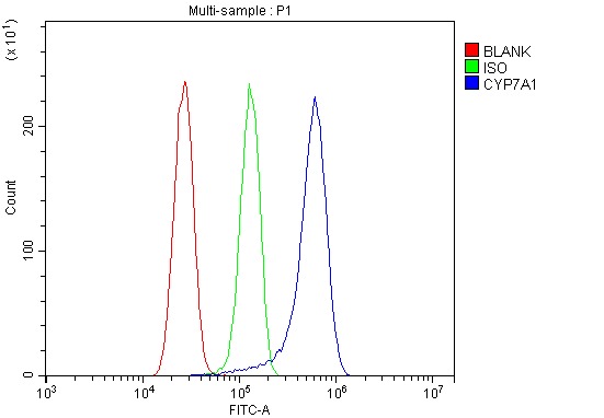
Supportive validation
- Submitted by
- Boster Biological Technology (provider)
- Main image
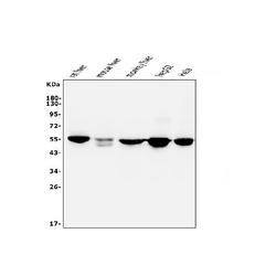
- Experimental details
- Western blot analysis of CYP7A1 using anti-CYP7A1 antibody (A01601). Electrophoresis was performed on a 5-20% SDS-PAGE gel at 70V (Stacking gel) / 90V (Resolving gel) for 2-3 hours. The sample well of each lane was loaded with 50ug of sample under reducing conditions. Lane 1: rat liver tissue lysates, Lane 2: mouse liver tissue lysates, Lane 3: monkey liver tissue lysates, Lane 4: human HepG2 whole cell lysates, Lane 5: human Hele whole cell lysates. After Electrophoresis, proteins were transferred to a Nitrocellulose membrane at 150mA for 50-90 minutes. Blocked the membrane with 5% Non-fat Milk/ TBS for 1.5 hour at RT. The membrane was incubated with rabbit anti-CYP7A1 antigen affinity purified polyclonal antibody (Catalog # A01601) at 0.5 μg/mL overnight at 4°C, then washed with TBS-0.1%Tween 3 times with 5 minutes each and probed with a goat anti-rabbit IgG-HRP secondary antibody at a dilution of 1:5000 for 1.5 hour at RT. The signal is developed using an Enhanced Chemiluminescent detection (ECL) kit (Catalog # EK1002) with Tanon 5200 system. A specific band was detected for CYP7A1 at approximately 54KD. The expected band size for CYP7A1 is at 54KD.
- Additional image