Explore
 Validate
 Learn
Learn Immunocytochemistry
Antibody data
- Antibody Data
- Antigen structure
- References [3]
- Comments [0]
- Validations
- Immunocytochemistry [1]
Submit
Validation data
Reference
Comment
Report error
- Product number
- MAB1924 - Provider product page

- Provider
- Abnova Corporation
- Proper citation
- Abnova Corporation Cat#MAB1924, RRID:AB_1672434
- Product name
- CENPE monoclonal antibody, clone 1H12
- Antibody type
- Monoclonal
- Description
- Mouse monoclonal antibody raised against full length recombinant.
- Isotype
- IgG
- Antibody clone number
- 1H12
- Storage
- Store at 4°C. For long term storage store at -20°C.Aliquot to avoid repeated freezing and thawing.
Submitted references Characterization of the kinetochore binding domain of CENP-E reveals interactions with the kinetochore proteins CENP-F and hBUBR1.
CENP-E is a putative kinetochore motor that accumulates just before mitosis.
CENP-E, a novel human centromere-associated protein required for progression from metaphase to anaphase.
Chan GK, Schaar BT, Yen TJ
The Journal of cell biology 1998 Oct 5;143(1):49-63
The Journal of cell biology 1998 Oct 5;143(1):49-63
CENP-E is a putative kinetochore motor that accumulates just before mitosis.
Yen TJ, Li G, Schaar BT, Szilak I, Cleveland DW
Nature 1992 Oct 8;359(6395):536-9
Nature 1992 Oct 8;359(6395):536-9
CENP-E, a novel human centromere-associated protein required for progression from metaphase to anaphase.
Yen TJ, Compton DA, Wise D, Zinkowski RP, Brinkley BR, Earnshaw WC, Cleveland DW
The EMBO journal 1991 May;10(5):1245-54
The EMBO journal 1991 May;10(5):1245-54
No comments: Submit comment
Supportive validation
- Submitted by
- Abnova Corporation (provider)
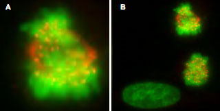
- Main image

- Experimental details
- Immunofluorescence microscopy of CENPE monoclonal antibody, clone 1H12 (Cat # MAB1924) was used to detect CENPE protein visible as discrete nuclear dots on prometaphase and metaphase cells that relocate to the spindle midzone at anaphase (panel A) .Interphase cells show no discrete staining (bottom left, panel B) .HeLa cells were fixed in paraformaldehyde and stained using this primary antibody.AlexaFluor 555 conjugated anti-Mouse antibody (red) was used for detection.DNA was stained using bis-benzimide (DAPI) (green) .Personal Communication, Tim Yen, Fox Chase Cancer Center, Philadelphia, PA.
- Validation comment
- Immunofluorescence