Explore
 Validate
 Learn
Learn Western blot
Antibody data
- Antibody Data
- Antigen structure
- References [1]
- Comments [0]
- Validations
- Western blot [1]
- Immunocytochemistry [1]
- Immunohistochemistry [2]
- Other assay [1]
Submit
Validation data
Reference
Comment
Report error
- Product number
- PA5-99403 - Provider product page

- Provider
- Invitrogen Antibodies
- Product name
- Aquaporin 5 Polyclonal Antibody
- Antibody type
- Polyclonal
- Antigen
- Synthetic peptide
- Description
- Antibody detects endogenous levels of total AQP5.
- Reactivity
- Human, Rat
- Host
- Rabbit
- Isotype
- IgG
- Vial size
- 100 µL
- Concentration
- 1 mg/mL
- Storage
- -20°C
Submitted references Organ-specific extracellular matrix directs trans-differentiation of mesenchymal stem cells and formation of salivary gland-like organoids in vivo.
Tran ON, Wang H, Li S, Malakhov A, Sun Y, Abdul Azees PA, Gonzalez AO, Cao B, Marinkovic M, Singh BB, Dean DD, Yeh CK, Chen XD
Stem cell research & therapy 2022 Jul 15;13(1):306
Stem cell research & therapy 2022 Jul 15;13(1):306
No comments: Submit comment
Supportive validation
- Submitted by
- Invitrogen Antibodies (provider)
- Main image

- Experimental details
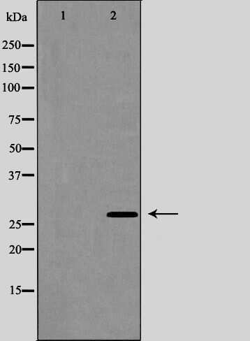
- Western blot analysis of Aquaporin 5 in HUVEC cell lysate (left lane: treated with the antigen-specific peptide). Samples were incubated with Aquaporin 5 polyclonal antibody (Product # PA5-99403).
Supportive validation
- Submitted by
- Invitrogen Antibodies (provider)
- Main image
- Experimental details
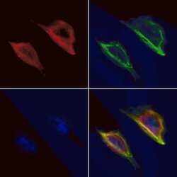
- Immunofluorescent analysis of Aquaporin 5 in HeLa cells. Samples were fixed with paraformaldehyde, permeabilized with 0.1% Triton X-100, blocked with 10% serum (45 min at 25°C), incubated with mouse anti-beta tubulin and Aquaporin 5 polyclonal antibody (Product # PA5-99403) using a dilution of 1:200 (1 hr, 37°C), and followed by goat anti-rabbit IgG Alexa Fluor 594 (red) and goat anti-mouse IgG Alexa Fluor 488 (green).
Supportive validation
- Submitted by
- Invitrogen Antibodies (provider)
- Main image

- Experimental details
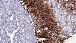
- Immunohistochemistry analysis of paraffin-embedded Aquaporin 5 in human lung tissue. Antigen retrieval was performed using citrate buffer. Samples were blocked with blocking buffer (1.5 hr, 22°C), incubated with Aquaporin 5 polyclonal antibody (Product # PA5-99403) using a dilution of 1:100 (1.5 hr, 22°C), followed by HRP conjugated goat anti-rabbit.
- Submitted by
- Invitrogen Antibodies (provider)
- Main image
- Experimental details
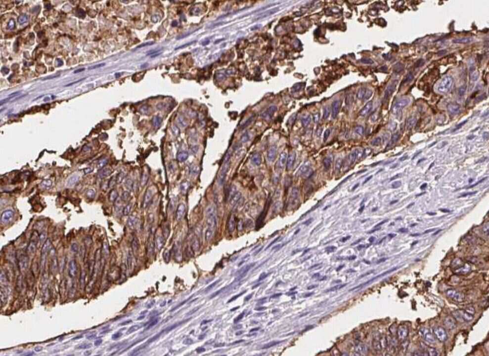
- Immunohistochemistry analysis of paraffin-embedded Aquaporin 5 in human urothelial cancer tissue. Antigen retrieval was performed using citrate buffer. Samples were blocked with blocking buffer (1.5 hr, 22°C), incubated with Aquaporin 5 polyclonal antibody (Product # PA5-99403) using a dilution of 1:100 (1.5 hr, 22°C), followed by HRP conjugated goat anti-rabbit.

Supportive validation
- Submitted by
- Invitrogen Antibodies (provider)
- Main image
- Experimental details
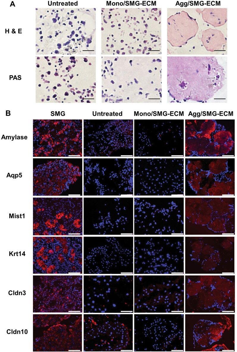
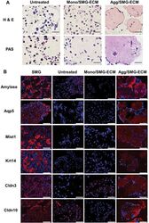
- Histological analysis of SMG-ECM-treated cultures reveals that aggregates display an expression pattern different from monolayer cells. A Paraffin sections of cells from 14 day cultures (i.e., untreated BM-MSCs [Untreated], SMG-ECM-treated aggregates [Agg/SMG-ECM], and monolayer [Mono/SMG-ECM] cells) were prepared and stained with H&E or PAS. PAS-positive cells are identified with arrows in the figure. Bar: 50 um. B Immunofluorescence staining of cells prepared as in A and rat SMG tissue (positive control) was used to evaluate the presence and localization of amylase, Aqp5, Mist1, Krt14, Cldn3, and Cldn10 protein. Staining with nonspecific isotype antibody was used as a negative control (not shown). Bar: 50 um