Explore
 Validate
 Learn
Learn Western blot
 Immunohistochemistry
Antibody data
- Antibody Data
- Antigen structure
- References [0]
- Comments [0]
- Validations
- Western blot [4]
- Immunocytochemistry [1]
Submit
Validation data
Reference
Comment
Report error
- Product number
- PA5-27971 - Provider product page

- Provider
- Invitrogen Antibodies
- Product name
- Anti-GPD1 Polyclonal Antibody
- Antibody type
- Polyclonal
- Antigen
- Recombinant protein fragment
- Description
- Recommended positive controls: HepG2, mouse brain. Predicted reactivity: Mouse (96%), Rat (95%), Xenopus laevis (84%), Pig (94%), Bovine (92%). Store product as a concentrated solution. Centrifuge briefly prior to opening the vial.
- Reactivity
- Human, Mouse, Rat
- Host
- Rabbit
- Isotype
- IgG
- Vial size
- 100 µL
- Concentration
- 0.74 mg/mL
- Storage
- Store at 4°C short term. For long term storage, store at -20°C, avoiding freeze/thaw cycles.
No comments: Submit comment
Supportive validation
- Submitted by
- Invitrogen Antibodies (provider)
- Main image
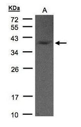
- Experimental details
- Western blot analysis of GPD1 using 50 µg of mouse brain lysate. Samples were loaded onto a 10% SDS-PAGE gel and probed with a GPD1 polyclonal antibody (Product # PA5-27971) at a dilution of 1:1000.

- Submitted by
- Invitrogen Antibodies (provider)
- Main image
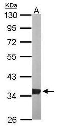
- Experimental details
- Western blot analysis of GPD1 using 30 µg of HepG2 lysate. Samples were loaded onto a 12% SDS-PAGE gel and probed with a GPD1 polyclonal antibody (Product # PA5-27971) at a dilution of 1:1000.
- Submitted by
- Invitrogen Antibodies (provider)
- Main image
- Experimental details
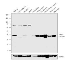
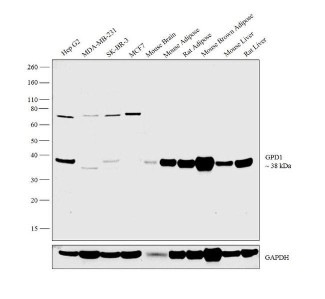
- Western blot analysis was performed on whole cell extracts (30 µg lysate) of HepG2 (Lane 1), MDA-MB-231 (Lane 2), SK-BR-3 (Lane 3), MCF7 (Lane 4), tissue extract Mouse Brain (Lane 5), Mouse Adipose (Lane 6), Rat Adipose (Lane 7), Mouse Brown Adipose (Lane 8), Mouse Liver (Lane 9) and Rat Liver (Lane 10). The blot was probed with Anti-GPD1 Polyclonal Antibody (Product # PA5-27971, 1:2000 dilution) and detected by chemiluminescence using Goat anti-Rabbit IgG (H+L) Superclonal™ Secondary Antibody, HRP conjugate (Product # A27036, 0.25 µg/ml, 1:4000 dilution). A 38 kDa band corresponding to GPD1 was observed across the cell lines and tissues tested except in breast cancer cell lines which are reported to be negative for GPD1 expression.
- Submitted by
- Invitrogen Antibodies (provider)
- Main image
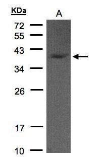
- Experimental details
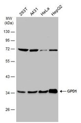
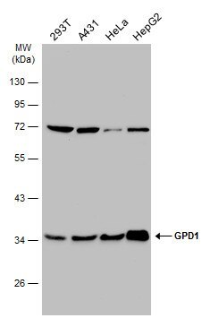
- Western Blot analysis of GPD1 was performed by separating 30 µg of various whole cell extracts by 10% SDS-PAGE. Proteins were transferred to a membrane and probed with a GPD1 Polyclonal Antibody (Product # PA5-27971) at a dilution of 1:1000 and a HRP-conjugated anti-rabbit IgG secondary antibody.
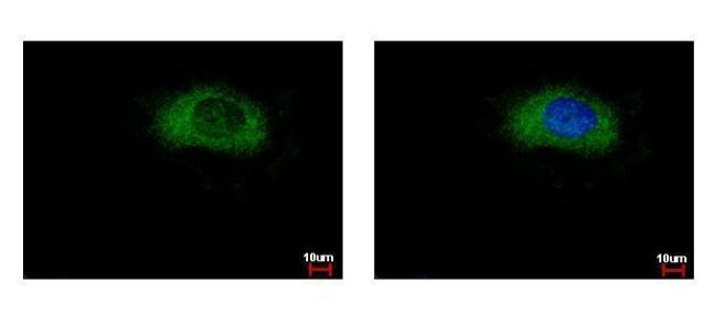
Supportive validation
- Submitted by
- Invitrogen Antibodies (provider)
- Main image
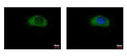
- Experimental details
- Immunofluorescent analysis of GPD1 showing staining in the cytoplasm of HeLa cells. HeLa cells were fixed in 4% paraformaldehyde at RT for 15 min and stained using a GPD1 polyclonal antibody (Product # PA5-27971) diluted at 1:500. Blue: Hoechst 33343 staining.