Explore
 Validate
 Learn
Learn Western blot
 Immunocytochemistry
Antibody data
- Antibody Data
- Antigen structure
- References [1]
- Comments [0]
- Validations
- Immunocytochemistry [6]
- Immunohistochemistry [1]
- Other assay [1]
Submit
Validation data
Reference
Comment
Report error
- Product number
- PA5-79747 - Provider product page

- Provider
- Invitrogen Antibodies
- Product name
- NOD1 Polyclonal Antibody
- Antibody type
- Polyclonal
- Antigen
- Recombinant full-length protein
- Description
- Reconstitute with 0.2 mL of distilled water to yield a concentration of 500 µg/mL. Positive Control - WB: A549 whole cell, Rat Cardiac Muscle Tissue. IHC: Human Placenta tissue. ICC/IF: A549 Cell, Hela Cell, SMMC-7721 Cell.
- Reactivity
- Human, Rat
- Host
- Rabbit
- Isotype
- IgG
- Vial size
- 100 μg
- Concentration
- 500 μg/mL
- Storage
- -20°C
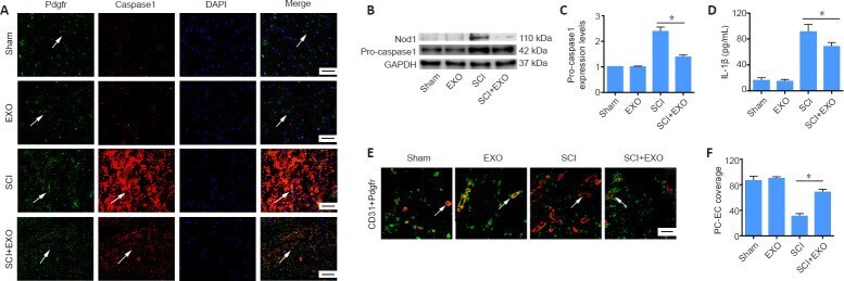
Submitted references Exosomes derived from bone marrow mesenchymal stem cells protect the injured spinal cord by inhibiting pericyte pyroptosis.
Zhou Y, Wen LL, Li YF, Wu KM, Duan RR, Yao YB, Jing LJ, Gong Z, Teng JF, Jia YJ
Neural regeneration research 2022 Jan;17(1):194-202
Neural regeneration research 2022 Jan;17(1):194-202
No comments: Submit comment
Supportive validation
- Submitted by
- Invitrogen Antibodies (provider)
- Main image

- Experimental details
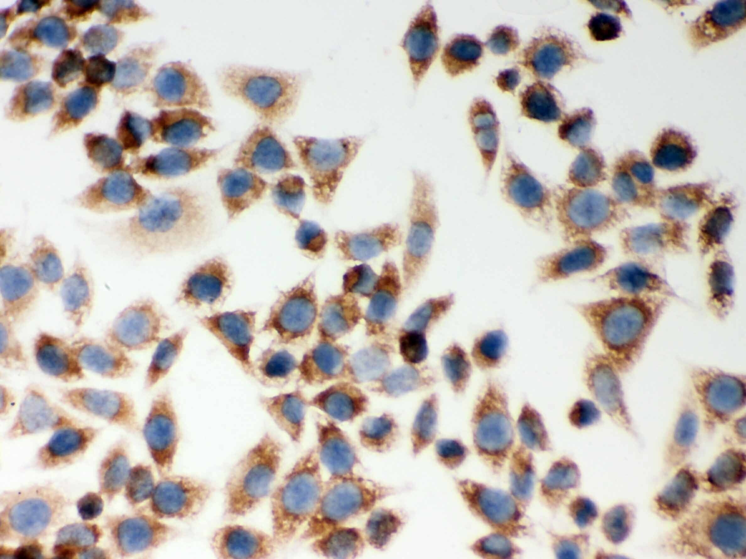
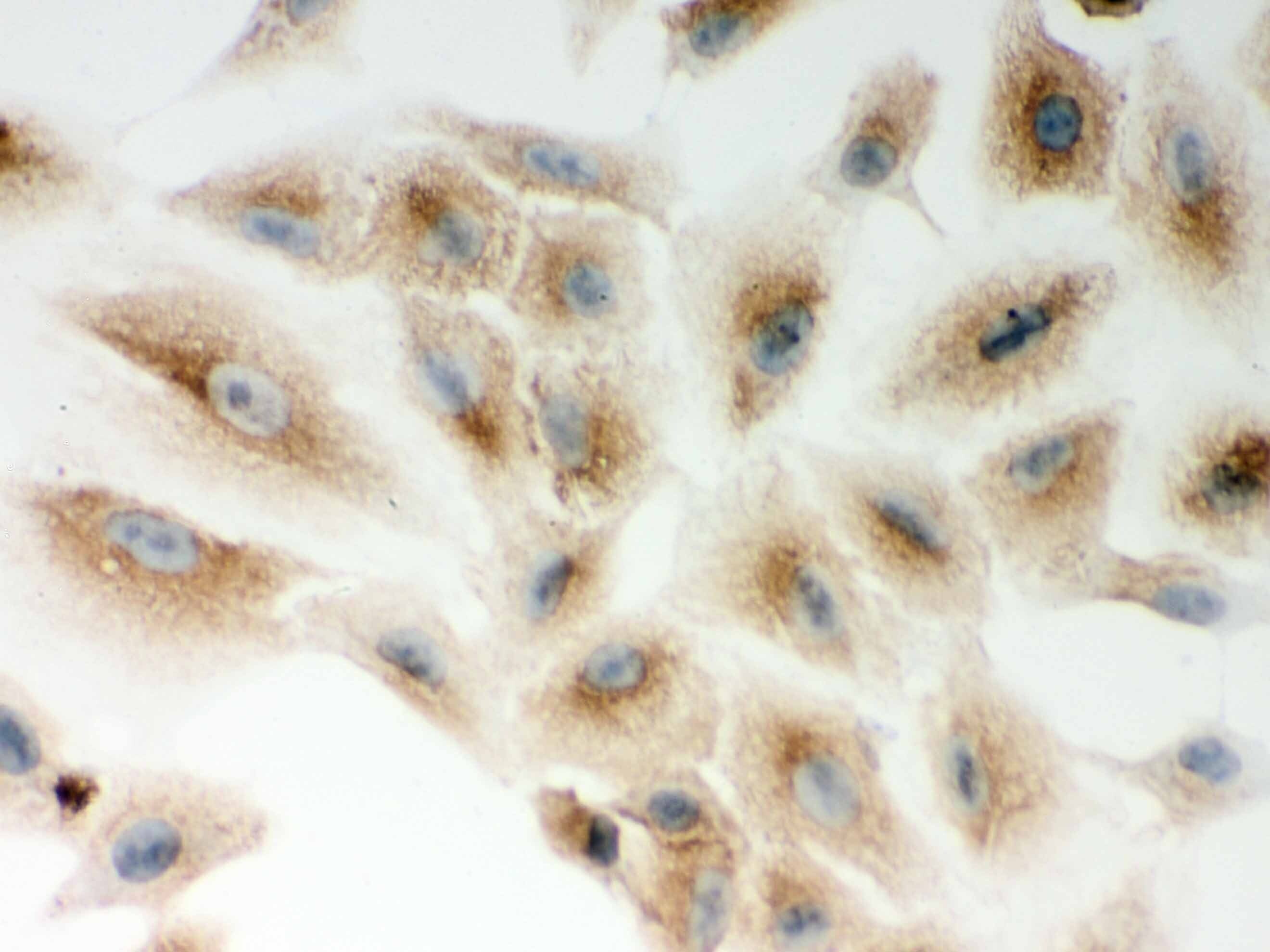
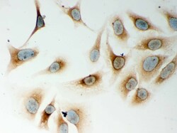
- Immunocytochemistry analysis of NOD1 on HeLa cells. Antigen retrieval was performed using citrate buffer (pH6, epitope retrieval solution) for 20 mins. Sample was blocked using 10% goat serum, incubated with NOD1 polyclonal antibody (Product# PA5-79747) with a dilution of 1 µg/mL (overnight at 4°C). Development was performed using Streptavidin-Biotin-Complex (SABC) with DAB chromogen method.

- Submitted by
- Invitrogen Antibodies (provider)
- Main image

- Experimental details
- Immunocytochemistry analysis of NOD1 on SMMC-7721 cells. Antigen retrieval was performed using citrate buffer (pH6, epitope retrieval solution) for 20 mins. Sample was blocked using 10% goat serum, incubated with NOD1 polyclonal antibody (Product# PA5-79747) with a dilution of 1 µg/mL (overnight at 4°C). Development was performed using Streptavidin-Biotin-Complex (SABC) with DAB chromogen method.
- Submitted by
- Invitrogen Antibodies (provider)
- Main image

- Experimental details
- Immunocytochemistry analysis of NOD1 on A549 cells. Antigen retrieval was performed using citrate buffer (pH6, epitope retrieval solution) for 20 mins. Sample was blocked using 10% goat serum, incubated with NOD1 polyclonal antibody (Product# PA5-79747) with a dilution of 1 µg/mL (overnight at 4°C). Development was performed using Streptavidin-Biotin-Complex (SABC) with DAB chromogen method.

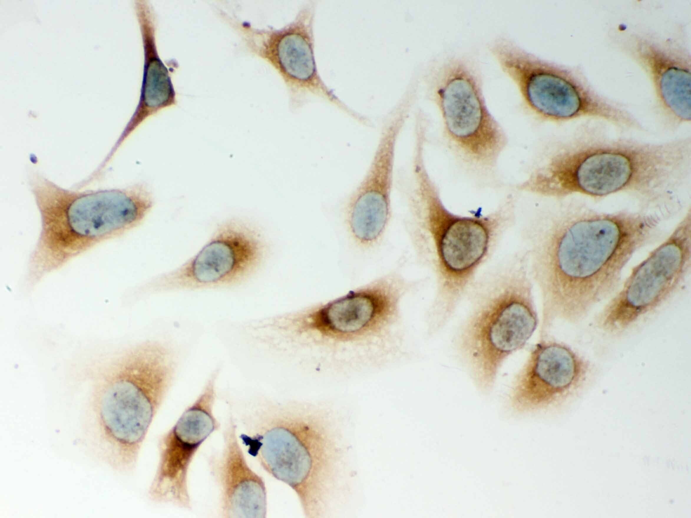
- Submitted by
- Invitrogen Antibodies (provider)
- Main image
- Experimental details
- Immunocytochemistry analysis of NOD1 on HeLa cells. Antigen retrieval was performed using citrate buffer (pH6, epitope retrieval solution) for 20 mins. Sample was blocked using 10% goat serum, incubated with NOD1 polyclonal antibody (Product# PA5-79747) with a dilution of 1 µg/mL (overnight at 4°C). Development was performed using Streptavidin-Biotin-Complex (SABC) with DAB chromogen method.
- Submitted by
- Invitrogen Antibodies (provider)
- Main image
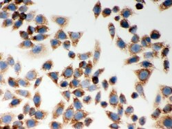
- Experimental details
- Immunocytochemistry analysis of NOD1 on SMMC-7721 cells. Antigen retrieval was performed using citrate buffer (pH6, epitope retrieval solution) for 20 mins. Sample was blocked using 10% goat serum, incubated with NOD1 polyclonal antibody (Product# PA5-79747) with a dilution of 1 µg/mL (overnight at 4°C). Development was performed using Streptavidin-Biotin-Complex (SABC) with DAB chromogen method.

Supportive validation
- Submitted by
- Invitrogen Antibodies (provider)
- Main image
- Experimental details
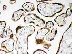
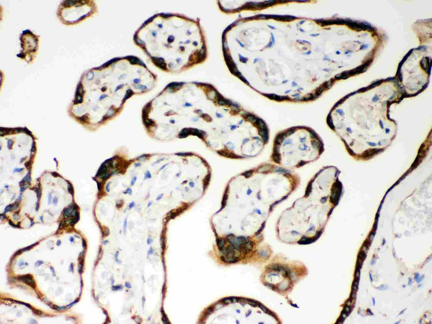
- Immunohistochemistry analysis of NOD1 on paraffin-embedded human placenta tissue. Antigen retrieval was performed using citrate buffer (pH6, epitope retrieval solution) for 20 mins. Sample was blocked using 10% goat serum, incubated with NOD1 polyclonal antibody (Product# PA5-79747) with a dilution of 1 µg/mL (overnight at 4°C), and followed by biotinylated goat anti-rabbit IgG (30 minutes at 37°C). Development was performed using Streptavidin-Biotin-Complex (SABC) with DAB chromogen method.
Supportive validation
- Submitted by
- Invitrogen Antibodies (provider)
- Main image
- Experimental details
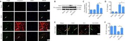
- Figure 4 BMSC-EXOs decrease pyroptosis in pericytes and increase pericyte coverage in vivo . (A) Colocalization of Pdgfr and caspase 1 in the spinal cord at 3 dpi. Arrows indicate pericytes. Red: DyLight 594, caspase 1; green: DyLight 488, Pdgfr; blue: DAPI. (B, C) Expression of Nod1 (data not shown) and pro-caspase 1 (fold-change compared with the Sham group) assessed by western blot assay. (D) Expression of IL-1beta assessed by enzyme-linked immunosorbent assay. (E) Representative immunofluorescence images of Pdgfrbeta + pericytes and CD31 + endothelial cells after SCI. Compared with the Sham and EXO groups, the SCI group showed loss of pericytes and low PC/EC coverage. Exosome treatment reduced these changes. Arrows indicate microvessels. Red: DyLight 594, CD31 + endothelial cells; green: DyLight 488, Pdgfrbeta+ pericytes; blue: DAPI. Nuclei were labeled with DAPI. Scale bars: 50 um in A, and 20 um in E. (F) Percentage of PC/EC coverage in spinal cord microvessels. Data are expressed as the mean +- SD ( n = 3 in C, n = 6 in D and F). * P < 0.05 (Student's t -test). BMSC-EXO: Bone mesenchymal stem cell-derived exosome; DAPI: 4',6-diamidino-2-phenylindole; EXO: exosome; PC/EC: pericyte/endothelial cell; Pdgfr: platelet-derived growth factor receptor; SCI: spinal cord injury.