Explore
 Validate
 Learn
Learn Western blot
Antibody data
- Antibody Data
- Antigen structure
- References [5]
- Comments [0]
- Validations
- Western blot [5]
- Immunocytochemistry [2]
- Immunohistochemistry [5]
- Other assay [3]
Submit
Validation data
Reference
Comment
Report error
- Product number
- PA5-52843 - Provider product page

- Provider
- Invitrogen Antibodies
- Product name
- TOMM20 Polyclonal Antibody
- Antibody type
- Polyclonal
- Antigen
- Recombinant full-length protein
- Description
- Immunogen sequence: KRRSDPNFKN RLRERRKKQK LAKERAGLSK LPDLKDAEAV QKFFLEEIQL GEELLAQGEY EKGVDHLTNA IAVCGQPQQL LQVLQQTLPP PVFQMLLTKL PTISQRIVSA QSLAEDDV Highest antigen sequence identity to the following orthologs: Mouse - 99%, Rat - 99%.
- Reactivity
- Human, Mouse
- Host
- Rabbit
- Isotype
- IgG
- Vial size
- 100 µL
- Concentration
- 0.2 mg/mL
- Storage
- Store at 4°C short term. For long term storage, store at -20°C, avoiding freeze/thaw cycles.
Submitted references The role of SQSTM1 (p62) in mitochondrial function and clearance in human cortical neurons.
Fitter Mitochondria Are Associated With Radioresistance in Human Head and Neck SQD9 Cancer Cells.
Nix-Mediated Mitophagy Modulates Mitochondrial Damage During Intestinal Inflammation.
Hypoxic Regulation of Mitochondrial Metabolism and Mitophagy in Nucleus Pulposus Cells Is Dependent on HIF-1α-BNIP3 Axis.
APP promotes osteoblast survival and bone formation by regulating mitochondrial function and preventing oxidative stress.
Poon A, Saini H, Sethi S, O'Sullivan GA, Plun-Favreau H, Wray S, Dawson LA, McCarthy JM
Stem cell reports 2021 May 11;16(5):1276-1289
Stem cell reports 2021 May 11;16(5):1276-1289
Fitter Mitochondria Are Associated With Radioresistance in Human Head and Neck SQD9 Cancer Cells.
Grasso D, Medeiros HCD, Zampieri LX, Bol V, Danhier P, van Gisbergen MW, Bouzin C, Brusa D, Grégoire V, Smeets H, Stassen APM, Dubois LJ, Lambin P, Dutreix M, Sonveaux P
Frontiers in pharmacology 2020;11:263
Frontiers in pharmacology 2020;11:263
Nix-Mediated Mitophagy Modulates Mitochondrial Damage During Intestinal Inflammation.
Vincent G, Novak EA, Siow VS, Cunningham KE, Griffith BD, Comerford TE, Mentrup HL, Stolz DB, Loughran P, Ranganathan S, Mollen KP
Antioxidants & redox signaling 2020 Jul 1;33(1):1-19
Antioxidants & redox signaling 2020 Jul 1;33(1):1-19
Hypoxic Regulation of Mitochondrial Metabolism and Mitophagy in Nucleus Pulposus Cells Is Dependent on HIF-1α-BNIP3 Axis.
Madhu V, Boneski PK, Silagi E, Qiu Y, Kurland I, Guntur AR, Shapiro IM, Risbud MV
Journal of bone and mineral research : the official journal of the American Society for Bone and Mineral Research 2020 Aug;35(8):1504-1524
Journal of bone and mineral research : the official journal of the American Society for Bone and Mineral Research 2020 Aug;35(8):1504-1524
APP promotes osteoblast survival and bone formation by regulating mitochondrial function and preventing oxidative stress.
Pan JX, Tang F, Xiong F, Xiong L, Zeng P, Wang B, Zhao K, Guo H, Shun C, Xia WF, Mei L, Xiong WC
Cell death & disease 2018 Oct 22;9(11):1077
Cell death & disease 2018 Oct 22;9(11):1077
No comments: Submit comment
Supportive validation
- Submitted by
- Invitrogen Antibodies (provider)
- Main image
- Experimental details
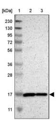
- Western blot analysis of TOMM20 in Lane 1: Marker (kDa) 230, 130, 95, 72, 56, 36, 28, 17, 11; Lane 2: Human cell line RT-4; Lane 3: Human cell line U-251MG sp. Samples were probed using a TOMM20 Polyclonal Antibody (Product # PA5-52843).

- Submitted by
- Invitrogen Antibodies (provider)
- Main image
- Experimental details
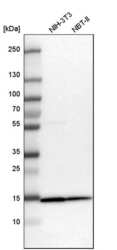
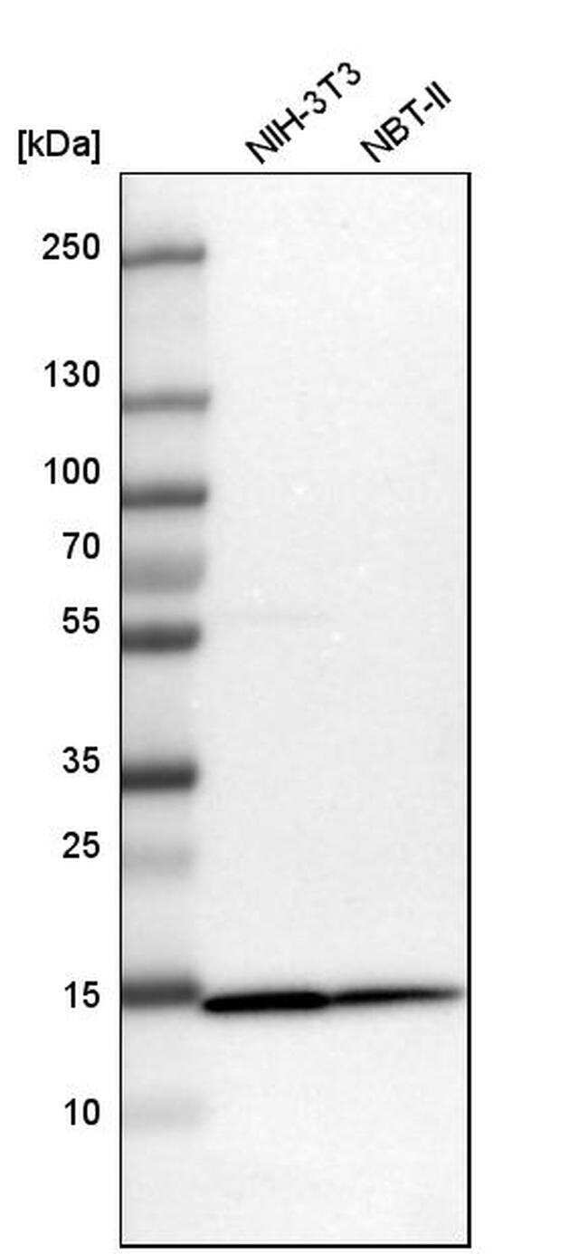
- Western blot analysis of TOMM20 in mouse cell line NIH-3T3 and rat cell line NBT-II using a TOMM20 Polyclonal Antibody (Product # PA5-52843).
- Submitted by
- Invitrogen Antibodies (provider)
- Main image
- Experimental details
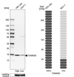
- Western blot analysis of TOMM20 in human cell lines HEK293 and MCF-7 using a TOMM20 Polyclonal Antibody (Product # PA5-52843). Corresponding TOMM20 RNA-seq data are presented for the same cell lines. Loading control: Anti-HSP90B1.

- Submitted by
- Invitrogen Antibodies (provider)
- Main image
- Experimental details
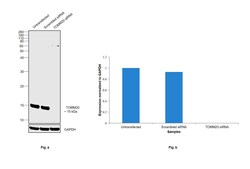
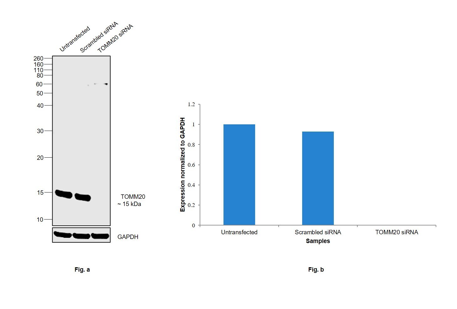
- Knockdown of TOMM20 was achieved by transfecting HeLa with TOMM20 specific siRNAs (Silencer® select Product # s18949, s18948). Western blot analysis (Fig. a) was performed using Membrane enriched extracts from the TOMM20 knockdown cells (lane 3), non-targeting scrambled siRNA transfected cells (lane 2) and untransfected cells (lane 1). The blot was probed with TOMM20 Polyclonal Antibody (Product # PA5-52843, 0.1 µg/mL) and Goat anti-Rabbit IgG (H+L) Superclonal™ Recombinant Secondary Antibody, HRP (Product # A27036, 1:4000 dilution). Densitometric analysis of this western blot is shown in histogram (Fig. b). Decrease in signal upon siRNA mediated knockdown confirms that antibody is specific to TOMM20.
- Submitted by
- Invitrogen Antibodies (provider)
- Main image

- Experimental details
- Western blot was performed using Anti-TOMM20 Polyclonal Antibody (Product # PA5-52843) and a 15 kDa band corresponding to TOMM20 was observed across cell lines and tissue extracts tested. Membrane enriched extracts (30 µg lysate) of HeLa (Lane 1), Hep G2 (Lane 2), MCF7 (Lane 3), HL-60 (Lane 4), NIH/3T3 (Lane 5), PC-12 (Lane 6) and tissue extracts of Mouse Brain (Lane 7) and Rat Brain (Lane 8) were electrophoresed using NuPAGE™ 12% Bis-Tris Protein Gel (Product # NP0342BOX). Resolved proteins were then transferred onto a Nitrocellulose membrane (Product # IB23001) by iBlot® 2 Dry Blotting System (Product # IB21001). The blot was probed with the primary antibody (0.1 µg/mL) and detected by chemiluminescence with Goat anti-Rabbit IgG (H+L) Superclonal™ Recombinant Secondary Antibody, HRP (Product # A27036, 1:4000 dilution) using the iBright FL 1000 (Product # A32752). Chemiluminescent detection was performed using Novex® ECL Chemiluminescent Substrate Reagent Kit (Product # WP20005).

Supportive validation
- Submitted by
- Invitrogen Antibodies (provider)
- Main image

- Experimental details
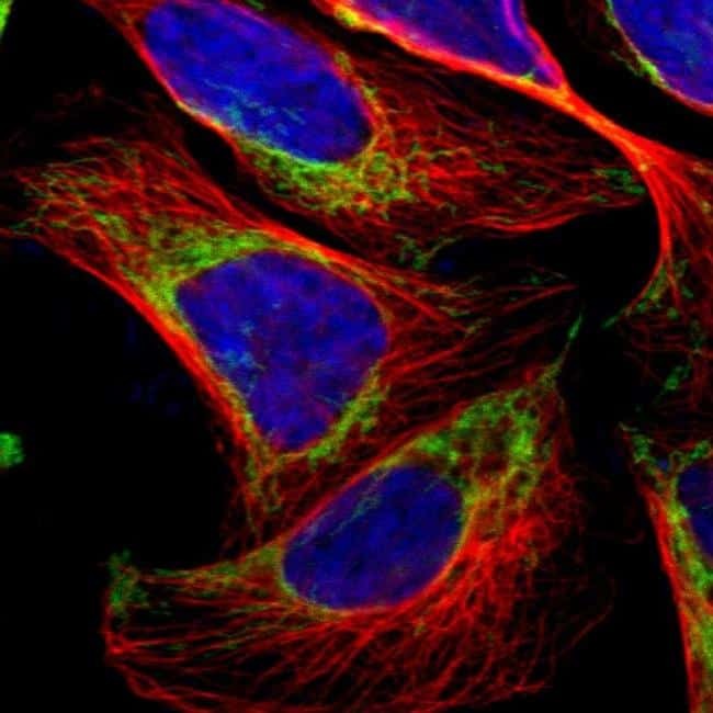
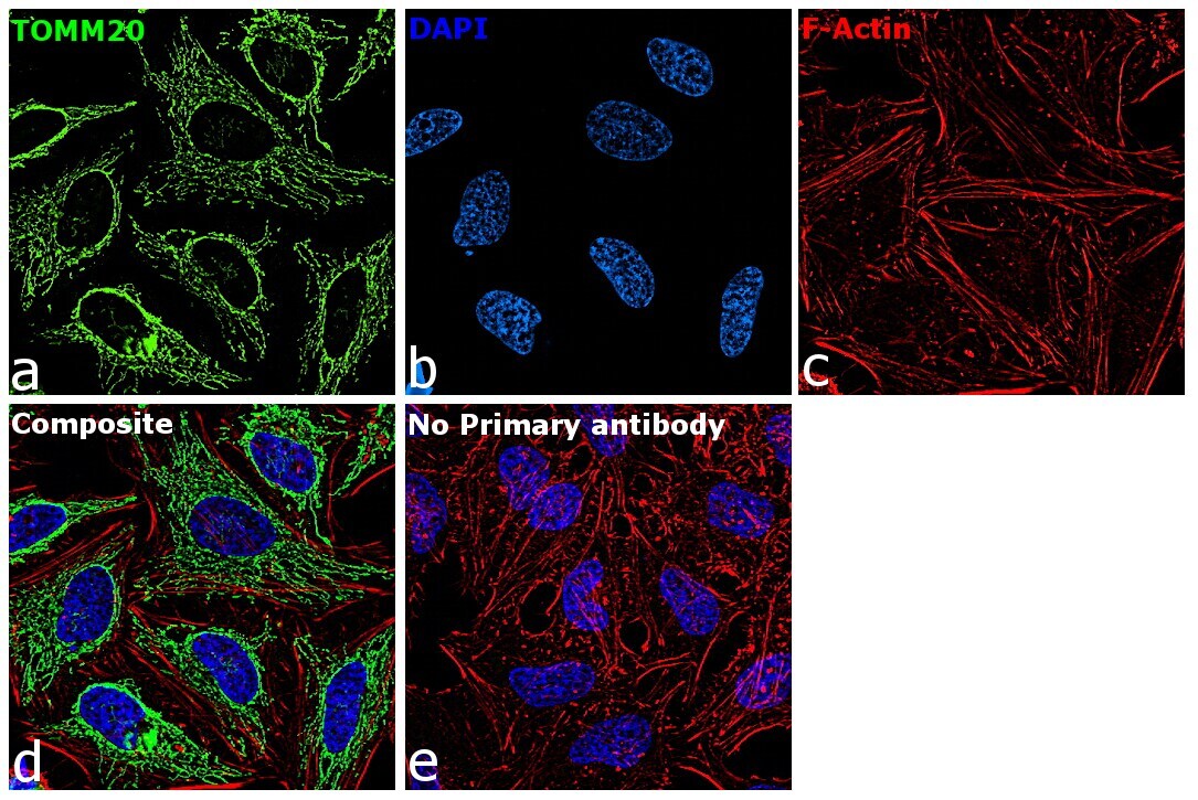
- Immunofluorescent staining of TOMM20 in human cell line U-2 OS shows positivity in mitochondria. Samples were probed using a TOMM20 Polyclonal Antibody (Product # PA5-52843).
- Submitted by
- Invitrogen Antibodies (provider)
- Main image

- Experimental details
- Immunofluorescence analysis of TOMM20 was performed using 70% confluent log phase HeLa cells. The cells were fixed with 4% paraformaldehyde for 10 minutes, permeabilized with 0.1% Triton™ X-100 for 15 minutes, and blocked with 2% BSA for 45 minutes at room temperature. The cells were labeled with TOMM20 Polyclonal Antibody (Product # PA5-52843) at 2 µg/mL in 0.1% BSA, incubated at 4 degree celsius overnight and then labeled with Donkey anti-Rabbit IgG (H+L) Highly Cross-Adsorbed Secondary Antibody, Alexa Fluor Plus 488 (Product # A32790), (1:2000), for 45 minutes at room temperature (Panel a: Green). Nuclei (Panel b:Blue) were stained with ProLong™ Diamond Antifade Mountant with DAPI (Product # P36962). F-actin (Panel c: Red) was stained with Rhodamine Phalloidin (Product # R415, 1:300). Panel d represents the merged image showing cytoplasmic(mitochondrial like pattern) localization. Panel e represents control cells with no primary antibody to assess background. The images were captured at 60X magnification.
Supportive validation
- Submitted by
- Invitrogen Antibodies (provider)
- Main image

- Experimental details
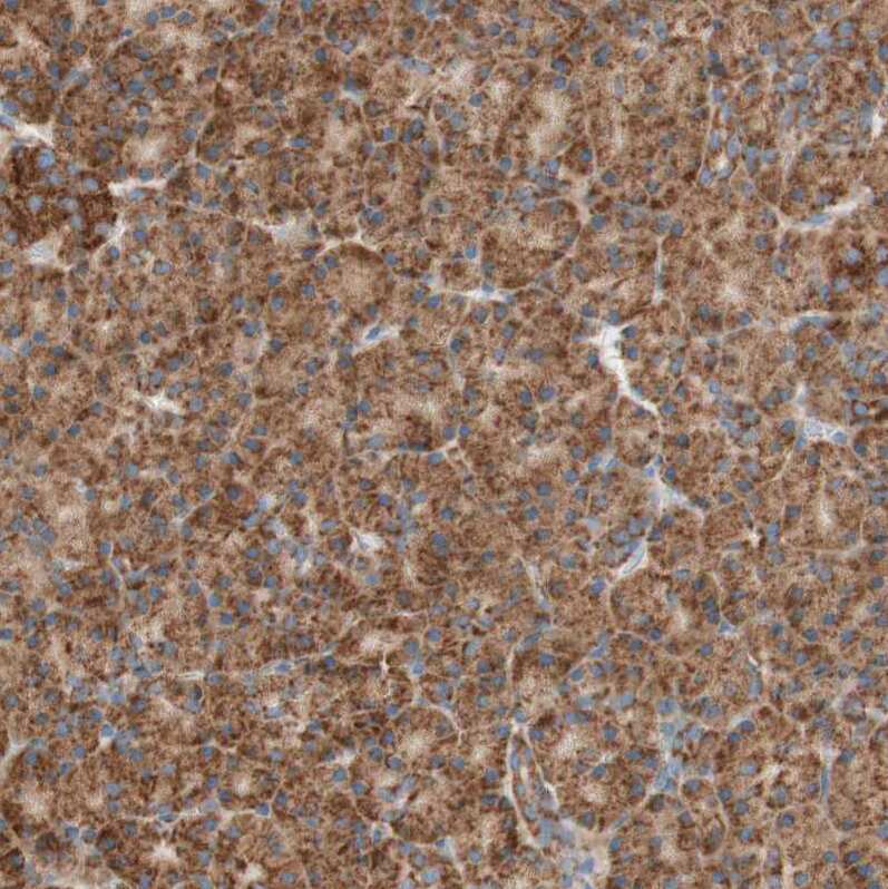
- Immunohistochemical staining of TOMM20 in human pancreas using TOMM20 Polyclonal Antibody (Product # PA5-52843) shows moderate to strong positivity in mitochondria in exocrine glandular cells.
- Submitted by
- Invitrogen Antibodies (provider)
- Main image

- Experimental details
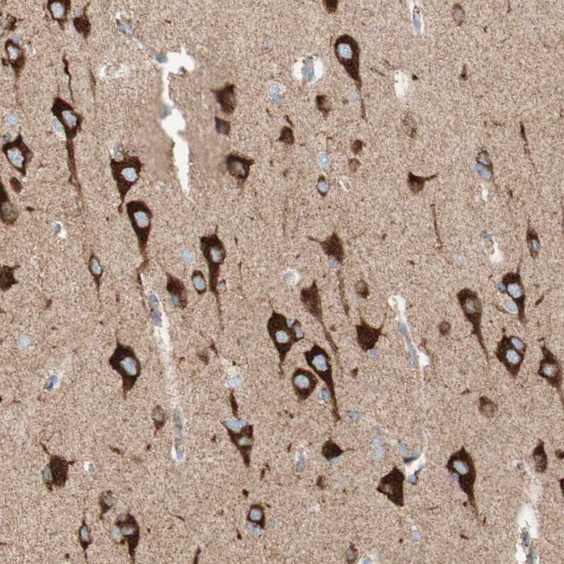
- Immunohistochemical staining of TOMM20 in human cerebral cortex using TOMM20 Polyclonal Antibody (Product # PA5-52843) shows moderate to strong positivity in mitochondria in neurons.
- Submitted by
- Invitrogen Antibodies (provider)
- Main image

- Experimental details
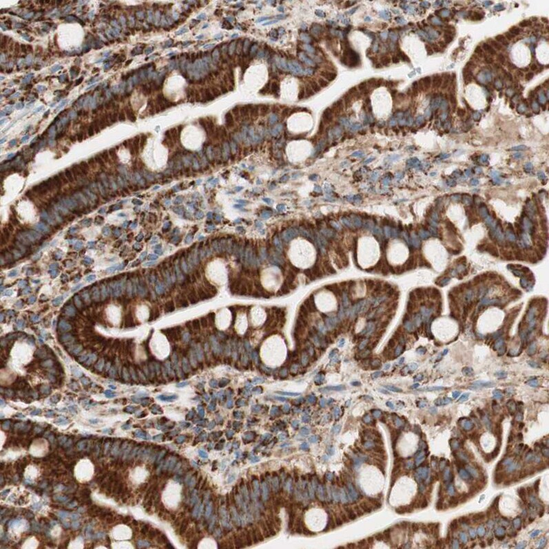
- Immunohistochemical staining of TOMM20 in human duodenum using TOMM20 Polyclonal Antibody (Product # PA5-52843) shows moderate to strong positivity in mitochondria in glandular cells.
- Submitted by
- Invitrogen Antibodies (provider)
- Main image
- Experimental details
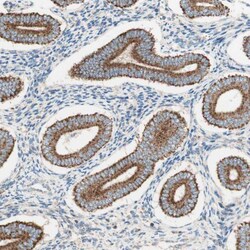
- Immunohistochemical staining of TOMM20 in human endometrium using TOMM20 Polyclonal Antibody (Product # PA5-52843) shows moderate to strong positivity in mitochondria in glandular cells.

- Submitted by
- Invitrogen Antibodies (provider)
- Main image

- Experimental details
- Immunohistochemical staining of TOMM20 in human prostate using TOMM20 Polyclonal Antibody (Product # PA5-52843) shows moderate to strong positivity in mitochondria in glandular cells.

Supportive validation
- Submitted by
- Invitrogen Antibodies (provider)
- Main image
- Experimental details
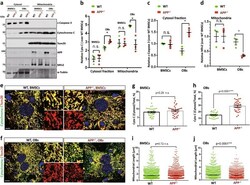
- Fig. 5 Increased cytochrome C release and mitochondrial fragmentation in APP -/- OBs, but not BMSCs. a - d Reduced mitochondrial cytochrome C and Mfn2 in APP -/- OBs, but not BMSCs. Mitochondrial and cytosol fractions of BMSCs and OBs from WT and APP -/- mice (2-M old) were subjected to Western bot analysis. a, Representative blots; b - d Quantifications of protein levels (compared with WT BMSCs); Data presented were mean +- SEM ( n = 3 mice/genotype); * p < 0.05. e - j Increased cytosolic cytochrome C and mitochondrial fragmentation in APP -/- OBs, but not BMSCs. OBs (D14 culture) were in vitro differentiated from BMSCs from WT and APP -/- mice (2-M old). Both BMSCs and OBs were co-immunostained with indicated antibodies. Mitochondria were labeled by Tom20. Representative images were shown in e , f . Higher power views of mitochondria (Tom20) and cytochrome C were also included in e , f Scale bar, 5 mum. g , h Quantifications of cytochrome C fluorescence in cytosol over total. The cytosolic cytochrome C was defined by cytochrome C + Tom20 - staining. Data presented were mean +- SEM ( n = 30 cells from 3-different cultures). i , j Quantifications of mitochondrial length. Shown were grouped column scatter (with mean +- SEM). n = 1500-2000 mitochondria from 30 different cells of each group. *** p < 0.0001

- Submitted by
- Invitrogen Antibodies (provider)
- Main image
- Experimental details
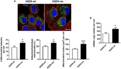
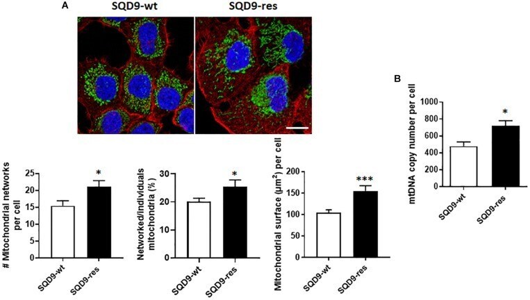
- FIGURE 10 Mitochondrial abundance and morphology differ between radiosensitive and radioresistant SQD9 cells. (A) Mitochondria were labeled with an anti-Tom20 antibody (green), the cytoskeleton with an anti-actin antibody (red) and cell nuclei with DAPI (blue). Representative pictures are shown on top (bar = 20 mum), and graphs show the number of mitochondrial networks per cell ( left ), the ratio of networked/individual mitochondria ( middle ) and the relative surface of cells occupied by mitochondria (MitoFootprint, right ) ( n = 20-34). (B) mtDNA content normalized to nuclear DNA content ( n = 8). * P < 0.05, *** P < 0.005 by Student''s t- test.
- Submitted by
- Invitrogen Antibodies (provider)
- Main image
- Experimental details
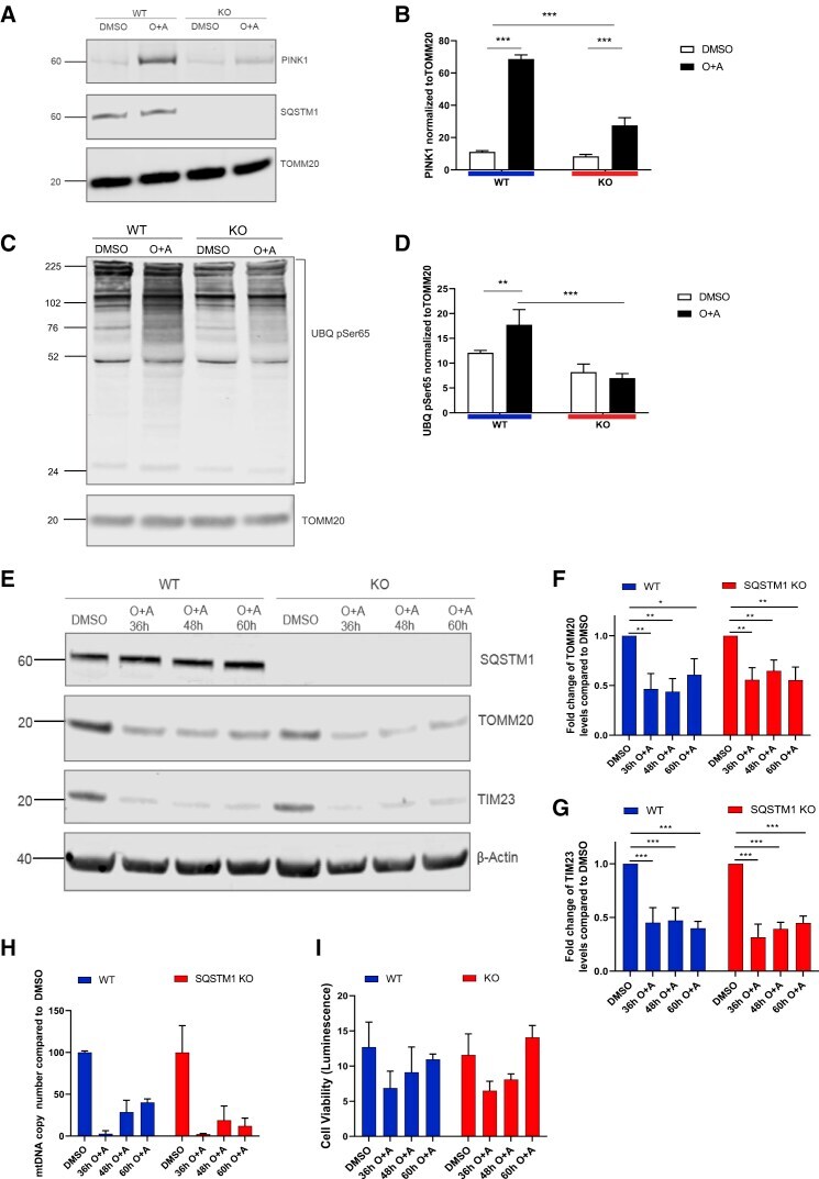
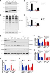
- Figure 4 SQSTM1 is not required for mitophagy in iPSC-derived cortical neurons (A) Immunoblots probed for mitochondrial components PINK1, SQSTM1, and TOMM20 after cells were treated with 1 muM oligomycin and antimycin A. (B) Bar graph displays quantified images showing PINK1 normalized to TOMM20 (loading control). Data are expressed as mean +- SEM of three independent experiments. Statistical differences were tested by two-tailed t test. *** p < 0.001. (C) Immunoblot for Ser65 (UBQ pSer65) and TOMM20 (using the same samples as examined in A). (D) Bar graph displays quantified images showing UBQ pSer65 normalized to TOMM20 (loading control). Data are expressed as mean +- SEM of three independent experiments. Statistical differences were tested by two-tailed t test. *** p < 0.001. (E) Immunoblot for TOMM20, TIM23, SQSTM1, and beta-actin from neurons treated for 36-60 h with 1 muM oligomycin and 1 muM antimycin A (or DMSO as control). (F and G) Bar graphs display quantified images showing fold change versus DMSO control of TOMM20 and TIM23 versus beta-actin loading control. Data are expressed as mean +- SEM of three independent experiments. Statistical differences were tested by two-tailed t test. * p < 0.05, ** p < 0.01, *** p < 0.001. (H) Quantitative mitochondrial DNA copy number in neurons, treated for 36-60 h with 1 muM oligomycin and 1 muM antimycin A (or DMSO as control), as determined by real-time PCR. Data are expressed as the mean ratio of mitochondrial-to-nuclear gene